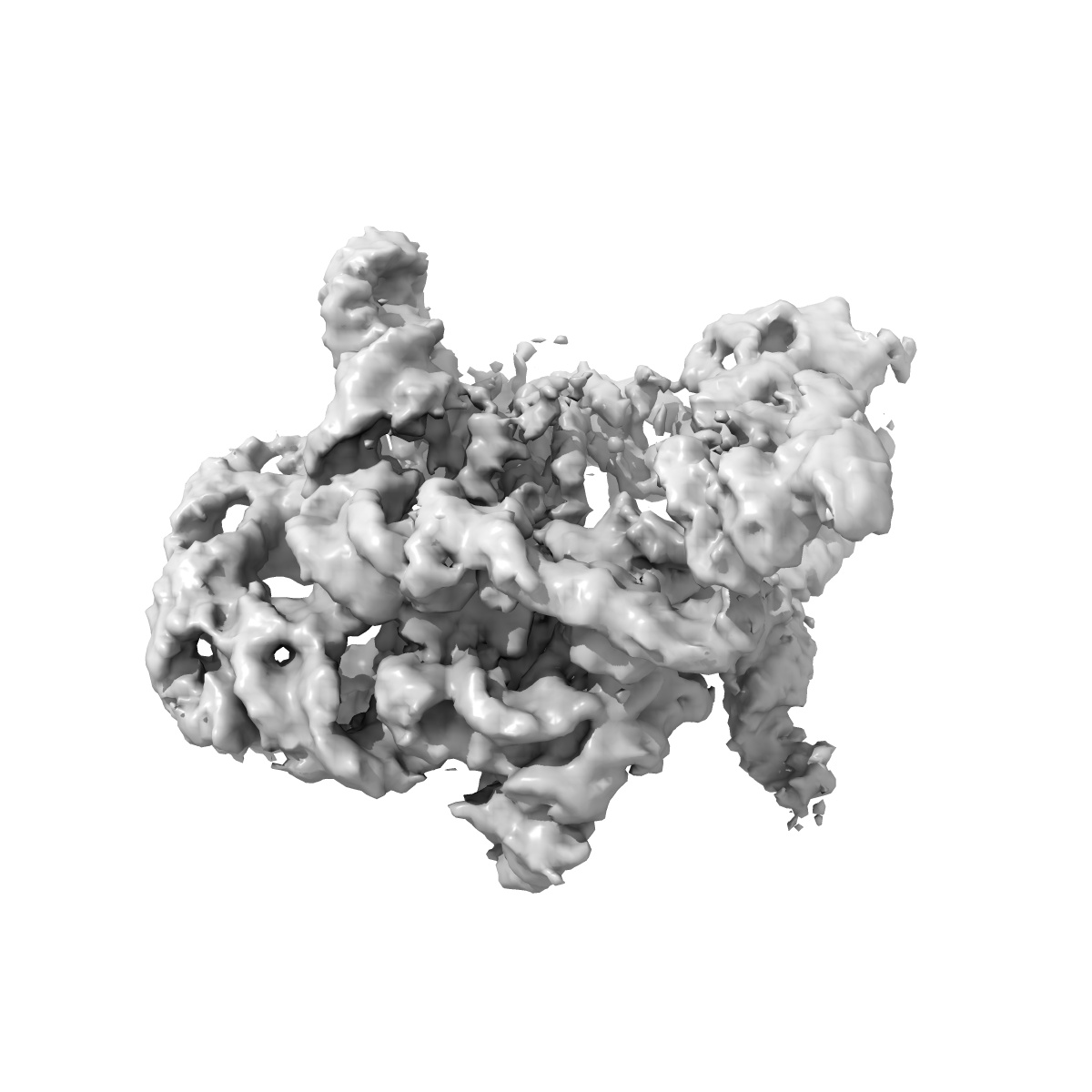
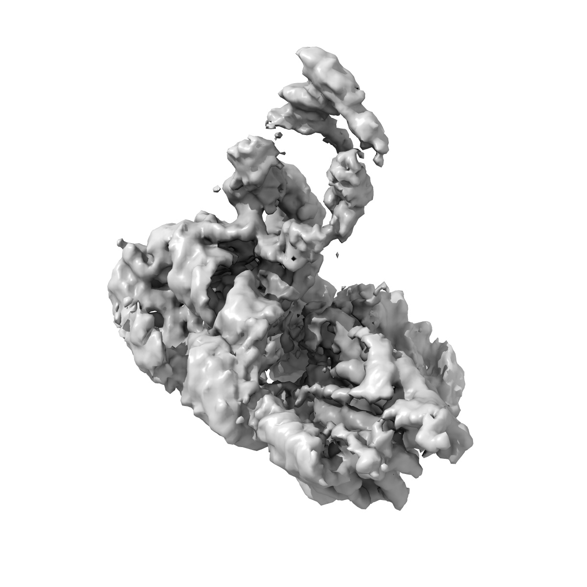
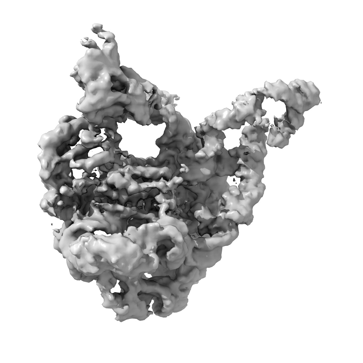
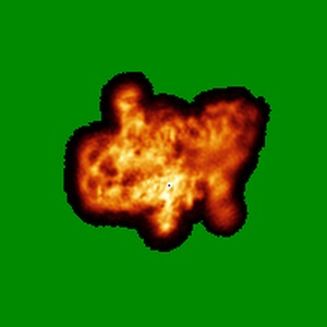
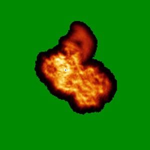
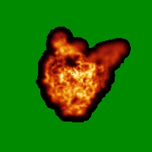
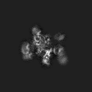
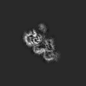
EMD-3333
Structure of a Group II Intron Complexed with its Reverse Transcriptase
EMD-3333
Single-particle4.2 Å
 Deposition: 14/02/2016
Map released: 04/05/2016
Last modified: 13/07/2016
Grid
Details: Quantifoil Cu-Rh R1.2/1.3 grid with thin carbon support
Vitrification
Microscope: FEI TITAN KRIOS
Illumination mode: FLOOD BEAM
Imaging mode: BRIGHT FIELD
Electron source: FIELD EMISSION GUN
Acceleration voltage: 300 kV
Nominal defocus: 1.2 µm - 3.0 µm
Specimen holder model: FEI TITAN KRIOS AUTOGRID HOLDER
Illumination mode: FLOOD BEAM
Imaging mode: BRIGHT FIELD
Electron source: FIELD EMISSION GUN
Acceleration voltage: 300 kV
Nominal defocus: 1.2 µm - 3.0 µm
Specimen holder model: FEI TITAN KRIOS AUTOGRID HOLDER
Image Recording
[1]
Final
reconstruction
Resolution: 4.2
Å
(
BY AUTHOR)
Resolution method: OTHER
Number of images used: 450296
Resolution method: OTHER
Number of images used: 450296
⌯ Applied Symmetry
Point group:
C1
Software
[1]
| Name | Version | Details |
|---|---|---|
| SPIDER, EMAN2, RELION | - | - |
CTF correction
Details: Each particle
Format: CCP4
Data type: IMAGE STORED AS FLOATING POINT NUMBER (4 BYTES)
Annotation details: Structure a of Group II Intron Complexed with its Reverse Transcriptase, Without Mask
Details: ::::EMDATABANK.org::::EMD-3333::::
Data type: IMAGE STORED AS FLOATING POINT NUMBER (4 BYTES)
Annotation details: Structure a of Group II Intron Complexed with its Reverse Transcriptase, Without Mask
Details: ::::EMDATABANK.org::::EMD-3333::::
⬡ Geometry
| X | Y | Z | |
|---|---|---|---|
| Origin | 0 | 0 | 0 |
| Dimensions (px) | 200 | 200 | 200 |
| Dimensions (Å) | 261.308 | 261.308 | 261.308 |
| Voxel size (Å) | 1.30654 | 1.30654 | 1.30654 |
Contour list
| Primary | Level | Source |
|---|---|---|
| True | 0.03 | AUTHOR |