EMD-3283
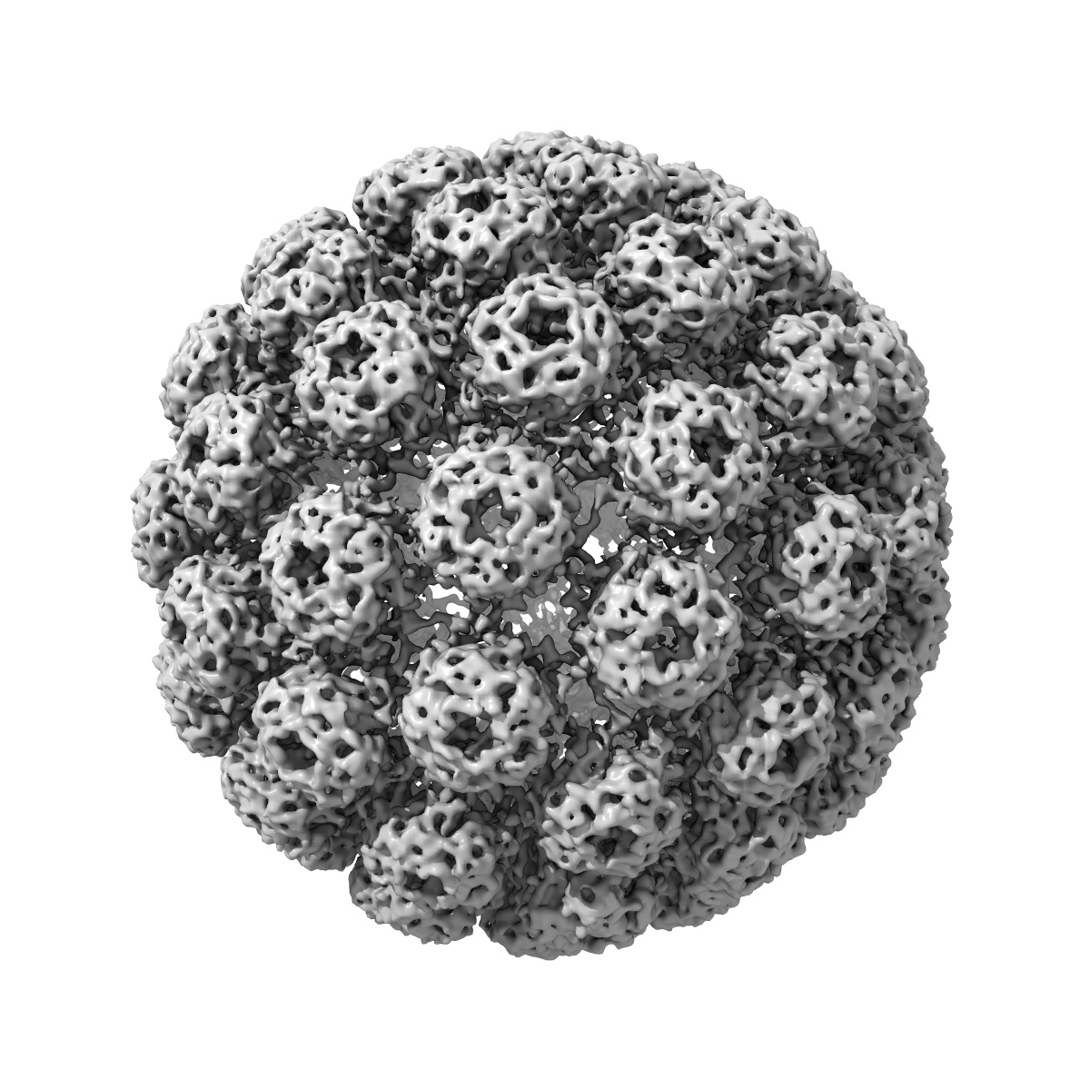
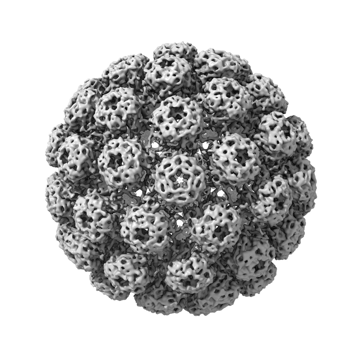
Cryo-EM structure of BK polyomavirus
EMD-3283
Single-particle7.6 Å
 Deposition: 17/12/2015
Map released: 27/04/2016
Last modified: 18/05/2016
Buffer
pH: 7.9
Details: 10mM HEPES pH 7.9, 1mM CaCl2, 1mM MgCl2, 5mM KCl
Details: 10mM HEPES pH 7.9, 1mM CaCl2, 1mM MgCl2, 5mM KCl
Grid
Details: Quantifoil R2/1 EM grids
Vitrification
Cryogen name: ETHANE
Chamber humidity: 100%
Instrument: FEI VITROBOT MARK IV
Method: 6.5 seconds blot before plunging
Chamber humidity: 100%
Instrument: FEI VITROBOT MARK IV
Method: 6.5 seconds blot before plunging
Microscope: FEI TECNAI F20
Illumination mode: FLOOD BEAM
Imaging mode: BRIGHT FIELD
Electron source: FIELD EMISSION GUN
Acceleration voltage: 200 kV
Nominal defocus: 0.6 µm - 4.8 µm
Nominal magnification: 19000.0
Specimen holder model: GATAN LIQUID NITROGEN
Minimum tilt angle: 0
Maximum tilt angle: 0
Illumination mode: FLOOD BEAM
Imaging mode: BRIGHT FIELD
Electron source: FIELD EMISSION GUN
Acceleration voltage: 200 kV
Nominal defocus: 0.6 µm - 4.8 µm
Nominal magnification: 19000.0
Specimen holder model: GATAN LIQUID NITROGEN
Minimum tilt angle: 0
Maximum tilt angle: 0
Image Recording
[1]
Detector category:
CCD
Detector model: GATAN K2 SUMMIT (4k x 4k)
Number of real images: 432
Average electron dose per image: 40 e/Å2
Details: 4 e-/A2/s, a 4 frames per second frame rate, and a 10 s exposure
Detector model: GATAN K2 SUMMIT (4k x 4k)
Number of real images: 432
Average electron dose per image: 40 e/Å2
Details: 4 e-/A2/s, a 4 frames per second frame rate, and a 10 s exposure
Format: CCP4
Data type: IMAGE STORED AS FLOATING POINT NUMBER (4 BYTES)
Annotation details: Reconstruction of BK polyomavirus (sharpened/masked)
Details: ::::EMDATABANK.org::::EMD-3283::::
Data type: IMAGE STORED AS FLOATING POINT NUMBER (4 BYTES)
Annotation details: Reconstruction of BK polyomavirus (sharpened/masked)
Details: ::::EMDATABANK.org::::EMD-3283::::
⬡ Geometry
| X | Y | Z | |
|---|---|---|---|
| Origin | -192 | -192 | -192 |
| Dimensions (px) | 384 | 384 | 384 |
| Dimensions (Å) | 737.27997 | 737.27997 | 737.27997 |
| Voxel size (Å) | 1.92 | 1.92 | 1.92 |
Contour list
| Primary | Level | Source |
|---|---|---|
| True | 0.022 | AUTHOR |