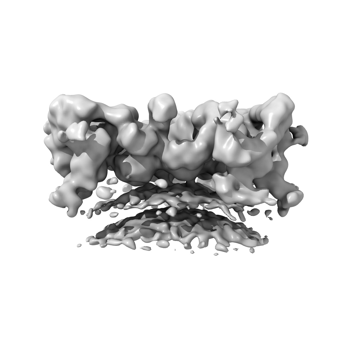
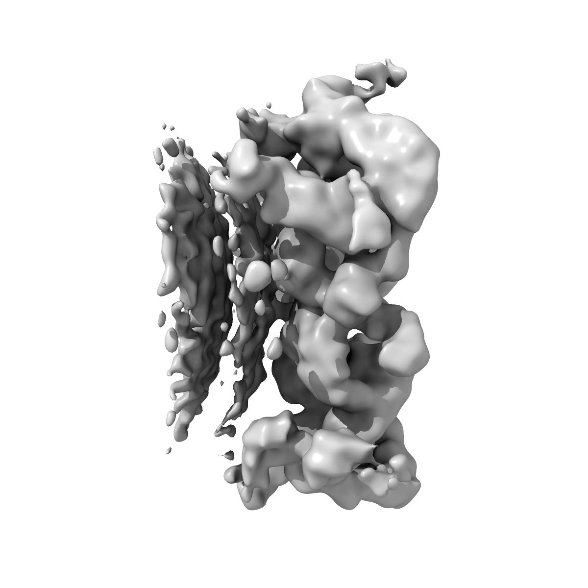
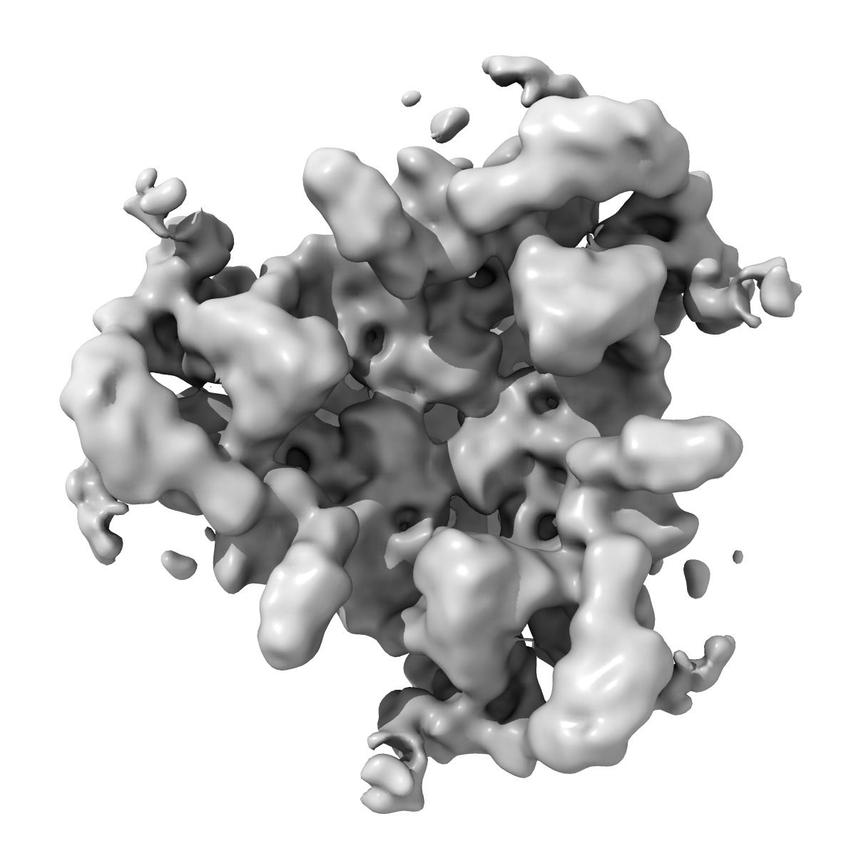
EMD-2985
The structure of the COPI coat triad
EMD-2985
Subtomogram averaging13.0 Å
 Deposition: 28/04/2015
Map released: 15/07/2015
Last modified: 22/07/2015
Name: COPI coat triad on the membrane
Name: COPI coat triad on the membrane
Number of unique components: 2
Details: The COPI coated vesicles were formed in an in vitro reconstituted budding reaction, which was plunge-frozen without further purification.
Number of unique components: 2
Details: The COPI coated vesicles were formed in an in vitro reconstituted budding reaction, which was plunge-frozen without further purification.
Molecular weight
| Experimental | Theoretical | Method |
|---|---|---|
| - | 1 MDa | - |
Name: Coat protein 1
(COPI)
UniProtKB PDBe-KB AlphaFold DB
UniProtKB PDBe-KB AlphaFold DB
Natural source
Molecular weight
| Experimental | Theoretical | Method |
|---|---|---|
| - | 560 kDa | - |
Recombinant Expression
| Organism | Strain | Cell | Plasmid |
|---|---|---|---|
| Spodoptera frugiperda | - | Sf9 | pFBDM |
Name: ADP-ribosylation factor 1
(Arf1)
UniProtKB PDBe-KB AlphaFold DB
UniProtKB PDBe-KB AlphaFold DB
Domains
Natural source
Molecular weight
| Experimental | Theoretical | Method |
|---|---|---|
| - | 20 kDa | - |
Recombinant Expression
| Organism | Strain | Cell | Plasmid |
|---|---|---|---|
| Escherichia coli BL21(DE3) | - | - | pOW12 |