EMD-2660
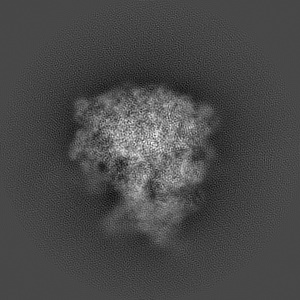
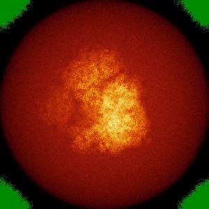
Cryo-EM structure of the Plasmodium falciparum 80S ribosome bound to the anti-protozoan drug emetine
EMD-2660
Single-particle3.2 Å
 Deposition: 22/05/2014
Map released: 18/06/2014
Last modified: 15/07/2015
Concentration: 0.6
mg/mL
Buffer
pH: 7.4
Details: 20 mM Hepes pH7.4, 40 mM KCH3COO, 10 mM NH4CH3COO, 10 mM Mg(CH3COO)2 and 5 mM 2-mecaptoethanol
Details: 20 mM Hepes pH7.4, 40 mM KCH3COO, 10 mM NH4CH3COO, 10 mM Mg(CH3COO)2 and 5 mM 2-mecaptoethanol
Staining
Type:
NEGATIVE
Details: Cryo-EM
Details: Cryo-EM
Grid
Details: 30 s on glow-discharged holey carbon grids (Quantifoil R2/2), onto which a home-made continuous carbon film
Vitrification
Cryogen name: ETHANE
Chamber humidity: 100%
Chamber temperature: 90 K
Instrument: FEI VITROBOT MARK IV
Method: Blot 2.5 seconds before plunging
Chamber humidity: 100%
Chamber temperature: 90 K
Instrument: FEI VITROBOT MARK IV
Method: Blot 2.5 seconds before plunging
Microscope: FEI POLARA 300
Illumination mode: FLOOD BEAM
Imaging mode: BRIGHT FIELD
Electron source: FIELD EMISSION GUN
Acceleration voltage: 300 kV
Nominal CS: 2 mm
Nominal defocus: 0.8 µm - 3.8 µm
Nominal magnification: 78000.0
Calibrated magnification: 104748.0
Specimen holder model: GATAN LIQUID NITROGEN
Alignment procedure: LEGACY (Astigmatism: Objective lens astigmatism was corrected at 78,000 times magnification, Electron beam tilt params: )
Temperature
Minimum: 80
K
Average: 85 K
Maximum: 90 K
Average: 85 K
Maximum: 90 K
Image Recording
[1]
Detector category:
CCD
Detector model: FEI FALCON II (4k x 4k)
Sampling interval: 14 µm
Number of real images: 1083
Average electron dose per image: 20 e/Å2
Details: Use a newly developed statistical movie processing approach to compensate for beam-induced movement.
Detector model: FEI FALCON II (4k x 4k)
Sampling interval: 14 µm
Number of real images: 1083
Average electron dose per image: 20 e/Å2
Details: Use a newly developed statistical movie processing approach to compensate for beam-induced movement.
Details: Use a newly developed statistical movie processing approach to compensate for beam-induced movement.
Final
reconstruction
Resolution: 3.2
Å
(
BY AUTHOR)
Resolution method: OTHER
Number of images used: 105247
Details: Use a newly developed statistical movie processing approach to compensate for beam-induced movement.
Resolution method: OTHER
Number of images used: 105247
Details: Use a newly developed statistical movie processing approach to compensate for beam-induced movement.
⌯ Applied Symmetry
Point group:
C1
Software
[1]
| Name | Version | Details |
|---|---|---|
| CTFFIND3, RELION | - | - |
CTF correction
Details: Each particle
Format: CCP4
Data type: IMAGE STORED AS FLOATING POINT NUMBER (4 BYTES)
Annotation details: Cryo-EM structure of the Plasmodium falciparum 80S ribosome bound to the anti-protozoan drug emetine
Details: ::::EMDATABANK.org::::EMD-2660::::
Data type: IMAGE STORED AS FLOATING POINT NUMBER (4 BYTES)
Annotation details: Cryo-EM structure of the Plasmodium falciparum 80S ribosome bound to the anti-protozoan drug emetine
Details: ::::EMDATABANK.org::::EMD-2660::::
⬡ Geometry
| X | Y | Z | |
|---|---|---|---|
| Origin | 0 | 0 | 0 |
| Dimensions (px) | 360 | 360 | 360 |
| Dimensions (Å) | 482.40002 | 482.40002 | 482.40002 |
| Voxel size (Å) | 1.34 | 1.34 | 1.34 |
Contour list
| Primary | Level | Source |
|---|---|---|
| True | 0.18 | AUTHOR |