







EMD-25536
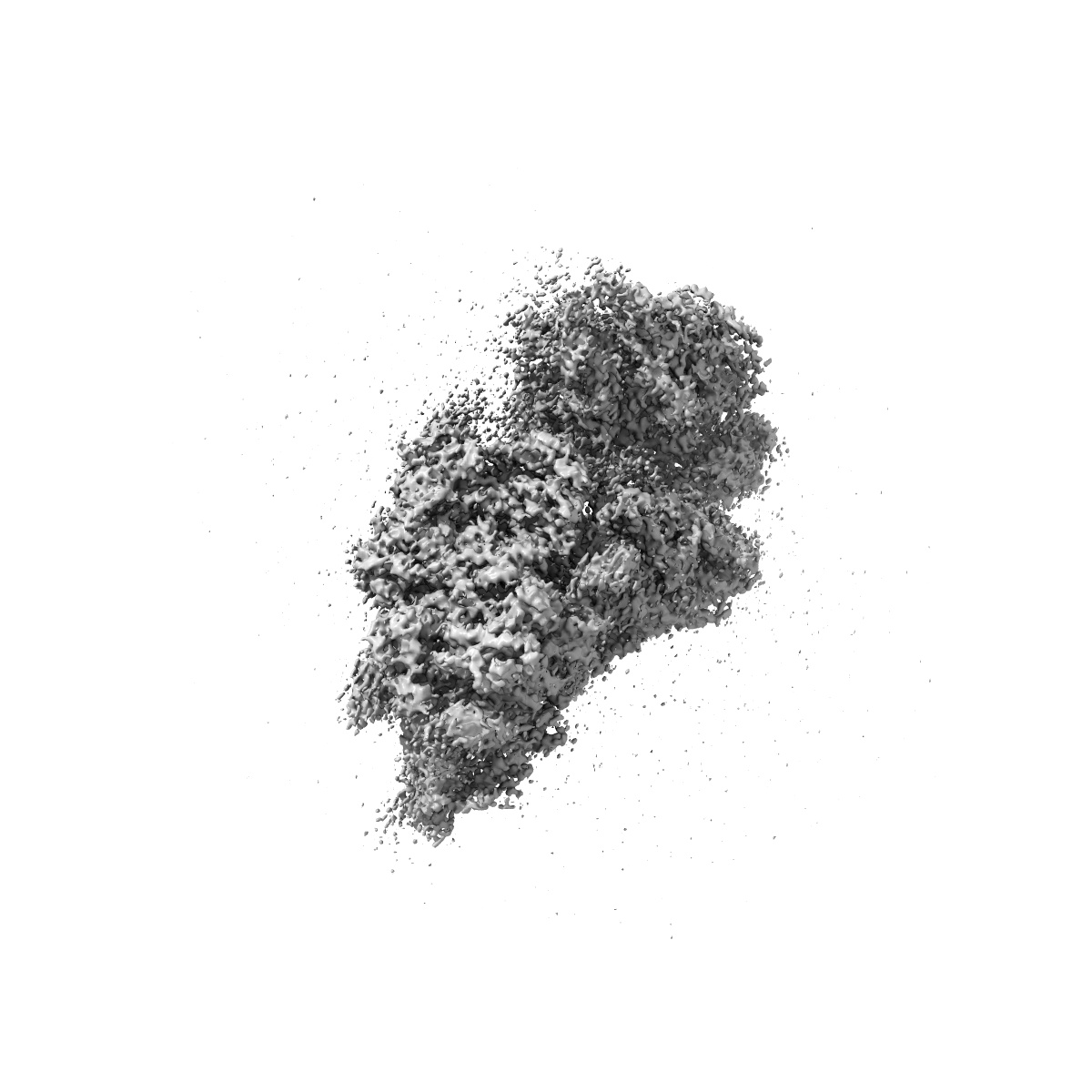
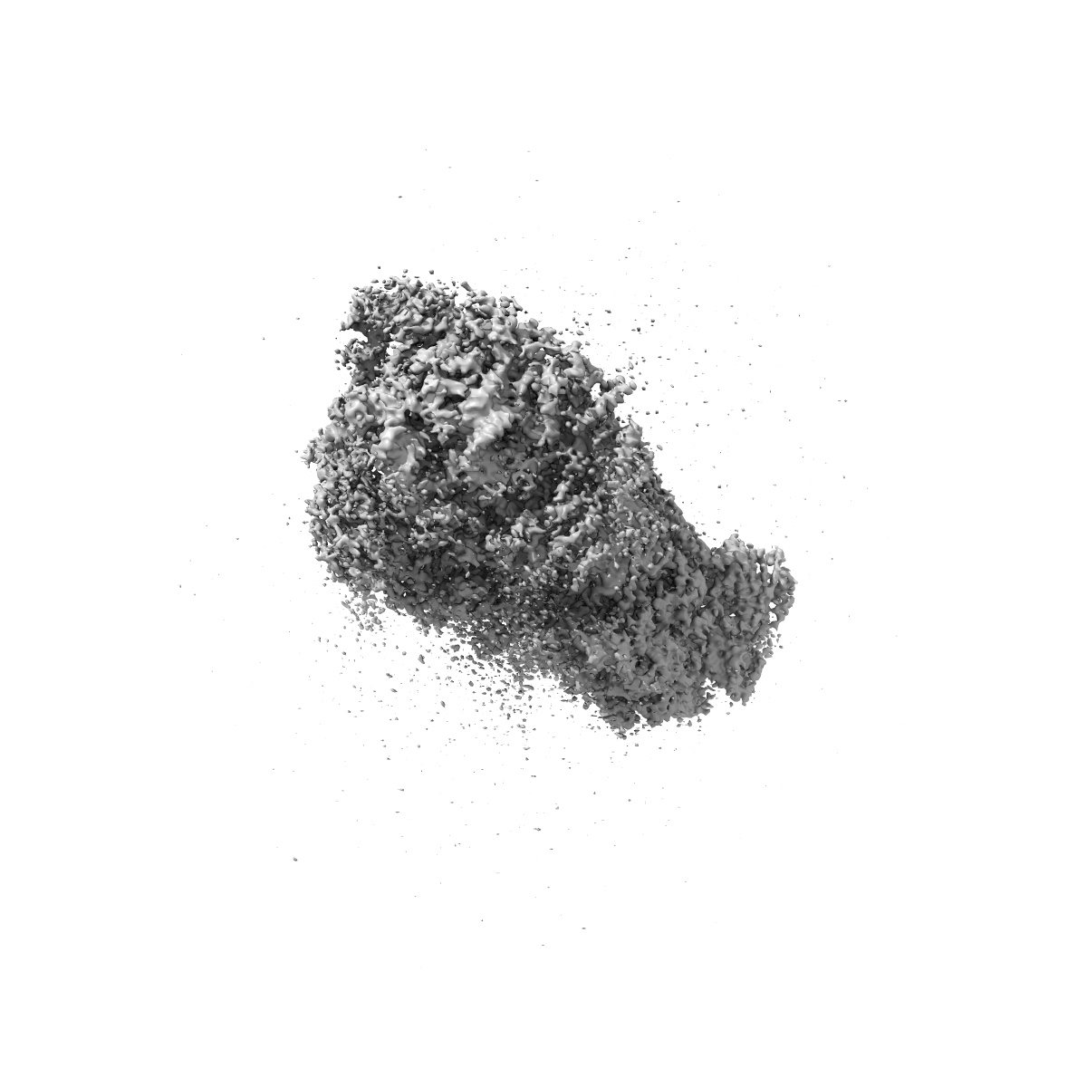
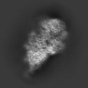
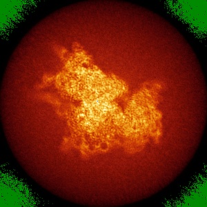
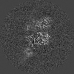
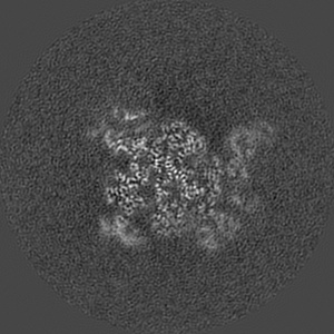
Structure of the wt IRES and 40S ribosome binary complex, open conformation. Structure 10(wt)
EMD-25536
Single-particle4.0 Å
 Deposition: 25/11/2021
Map released: 27/07/2022
Last modified: 16/10/2024
Sample Organism:
Oryctolagus cuniculus,
Hepatitis C virus (isolate 1)
Sample: 40S ribosomal small subunit with HCV IRES
Fitted models: 7syp
Deposition Authors: Brown ZP ,
Abaeva IS,
De S,
Hellen CUT ,
Pestova TV ,
Frank J
,
Abaeva IS,
De S,
Hellen CUT ,
Pestova TV ,
Frank J
Sample: 40S ribosomal small subunit with HCV IRES
Fitted models: 7syp
Deposition Authors: Brown ZP
 ,
Abaeva IS,
De S,
Hellen CUT ,
Pestova TV ,
Frank J
,
Abaeva IS,
De S,
Hellen CUT ,
Pestova TV ,
Frank J
Molecular architecture of 40S translation initiation complexes on the hepatitis C virus IRES.
Brown ZP ,
Abaeva IS,
De S,
Hellen CUT ,
Pestova TV ,
Frank J
(2022) EMBO J , 41 , e110581 - e110581
,
Abaeva IS,
De S,
Hellen CUT ,
Pestova TV ,
Frank J
(2022) EMBO J , 41 , e110581 - e110581
Grant Support:
- National Institutes of Health/National Institute of General Medical Sciences (NIH/NIGMS): R35 GM139453 (United States)
- National Institutes of Health/National Institute of General Medical Sciences (NIH/NIGMS): R01 GM55440 (United States)
- National Institutes of Health/National Institute of General Medical Sciences (NIH/NIGMS): 1R35GM122602 (United States)
- National Institutes of Health/National Institute Of Allergy and Infectious Diseases (NIH/NIAID): 1R01AI123406 (United States)