EMD-2379
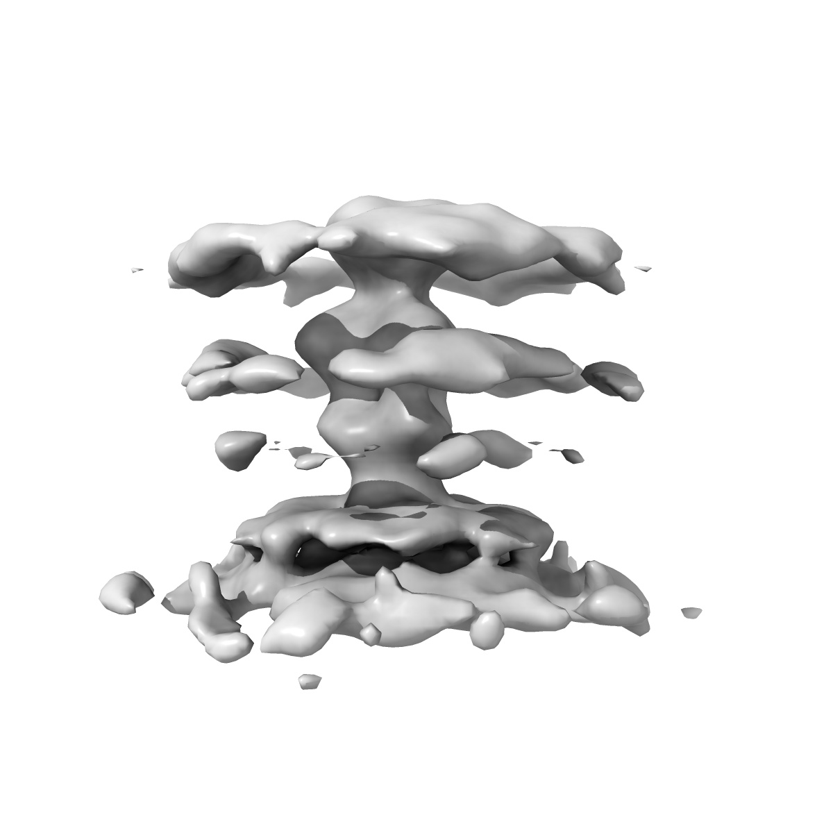
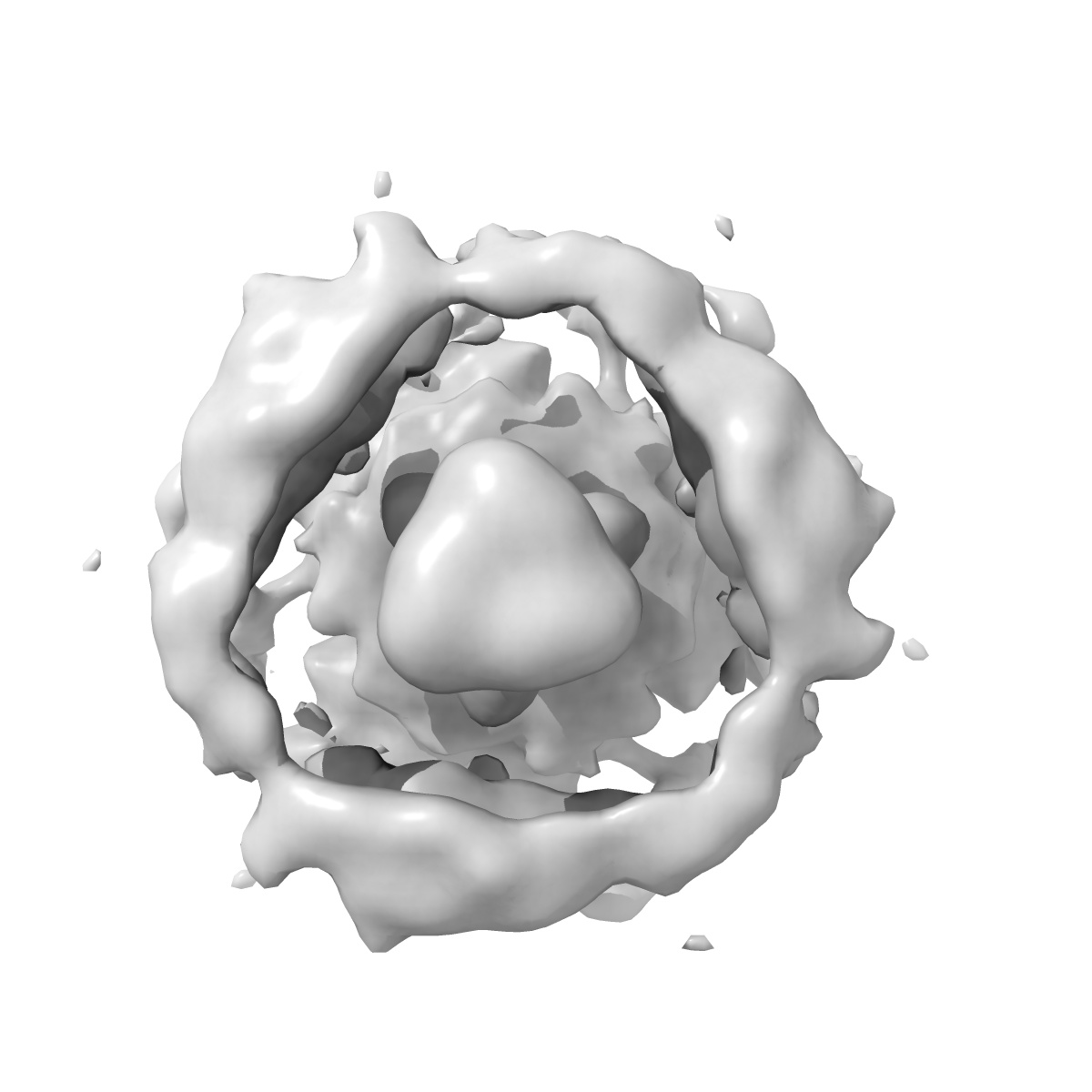
Structure of herpesvirus fusion glycoprotein B-bilayer complex revealing the protein-membrane and lateral protein-protein interaction
EMD-2379
Subtomogram averaging30.0 Å
 Deposition: 21/05/2013
Map released: 31/07/2013
Last modified: 21/08/2013
Concentration: 1.0
mg/mL
Buffer
pH: 5.5
Details: PBS with sodium citrate
Details: PBS with sodium citrate
Grid
Details: Cflat
Vitrification
Cryogen name: ETHANE-PROPANE MIXTURE
Chamber temperature: 120 K
Instrument: OTHER
Method: Blot manually for 3 s before plunging
Chamber temperature: 120 K
Instrument: OTHER
Method: Blot manually for 3 s before plunging
Microscope: FEI TECNAI F20
Illumination mode: FLOOD BEAM
Imaging mode: BRIGHT FIELD
Electron source: FIELD EMISSION GUN
Acceleration voltage: 200 kV
Nominal CS: 2 mm
Nominal defocus: 2.0 µm - 2.0 µm
Calibrated magnification: 67000.0
Specimen holder model: SIDE ENTRY, EUCENTRIC
Specimen holder details: Liquid nitrogen cooled
Illumination mode: FLOOD BEAM
Imaging mode: BRIGHT FIELD
Electron source: FIELD EMISSION GUN
Acceleration voltage: 200 kV
Nominal CS: 2 mm
Nominal defocus: 2.0 µm - 2.0 µm
Calibrated magnification: 67000.0
Specimen holder model: SIDE ENTRY, EUCENTRIC
Specimen holder details: Liquid nitrogen cooled
Image Recording
[1]
Detector category:
CCD
Detector model: GATAN ULTRASCAN 4000 (4k x 4k)
Sampling interval: 15 µm
Number of real images: 9
Average electron dose per image: 100 e/Å2
Bits per pixel: 12.0
Details: The dataset consists of 9 tomograms (containing 38 liposomes with bound gB). Data were binned by factor of 2.
Detector model: GATAN ULTRASCAN 4000 (4k x 4k)
Sampling interval: 15 µm
Number of real images: 9
Average electron dose per image: 100 e/Å2
Bits per pixel: 12.0
Details: The dataset consists of 9 tomograms (containing 38 liposomes with bound gB). Data were binned by factor of 2.
Tilt Series
[1]
| Axis 1 | Axis 2 | |||||
|---|---|---|---|---|---|---|
| Min. | Max. | Inc. | Min. | Max. | Inc. | Rotation |
| -60 ° | 60 ° | - | - | - | - | - |
Details: The sub-tomograms were picked manually from tomographic reconstructions of 38 liposomes
Final
reconstruction
Resolution: 30.0
Å
(
BY AUTHOR)
Resolution method: FSC 0.5 CUT-OFF
Algorithm: OTHER
Details: The best 730 spikes (of 996) were selected based on constrained cross correlation coefficient and by excluding overlaps. The long axis of the gB spike was assumed to be perpendicular to the plane of the membrane in the refinement.
Resolution method: FSC 0.5 CUT-OFF
Algorithm: OTHER
Details: The best 730 spikes (of 996) were selected based on constrained cross correlation coefficient and by excluding overlaps. The long axis of the gB spike was assumed to be perpendicular to the plane of the membrane in the refinement.
⌯ Applied Symmetry
Point group:
C3
Software
[1]
| Name | Version | Details |
|---|---|---|
| Jsubtomo | - | - |
CTF correction
Details: Low pass filter to the first zero crossing of the CTF
Format: CCP4
Data type: IMAGE STORED AS FLOATING POINT NUMBER (4 BYTES)
Annotation details: Subtomogram average of HSV-1 glycoprotein B bound to a lipid bilayer
Details: ::::EMDATABANK.org::::EMD-2379::::
Data type: IMAGE STORED AS FLOATING POINT NUMBER (4 BYTES)
Annotation details: Subtomogram average of HSV-1 glycoprotein B bound to a lipid bilayer
Details: ::::EMDATABANK.org::::EMD-2379::::
⬡ Geometry
| X | Y | Z | |
|---|---|---|---|
| Origin | -50 | -50 | -50 |
| Dimensions (px) | 100 | 100 | 100 |
| Dimensions (Å) | 460.0 | 460.0 | 460.0 |
| Voxel size (Å) | 4.6 | 4.6 | 4.6 |
Contour list
| Primary | Level | Source |
|---|---|---|
| True | 2.0 | AUTHOR |