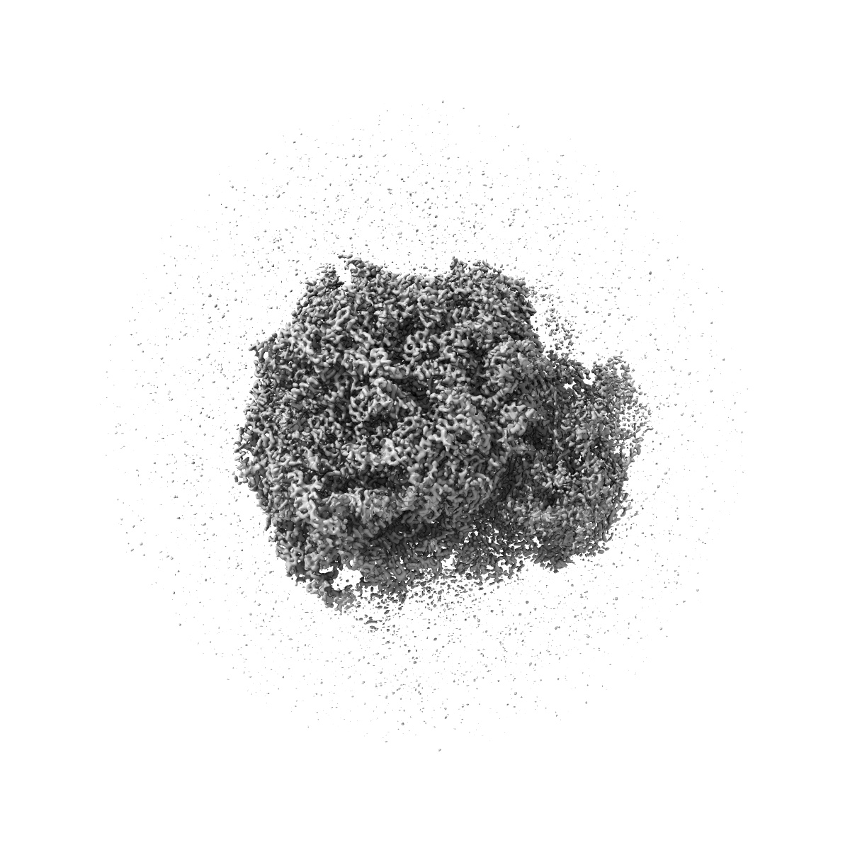
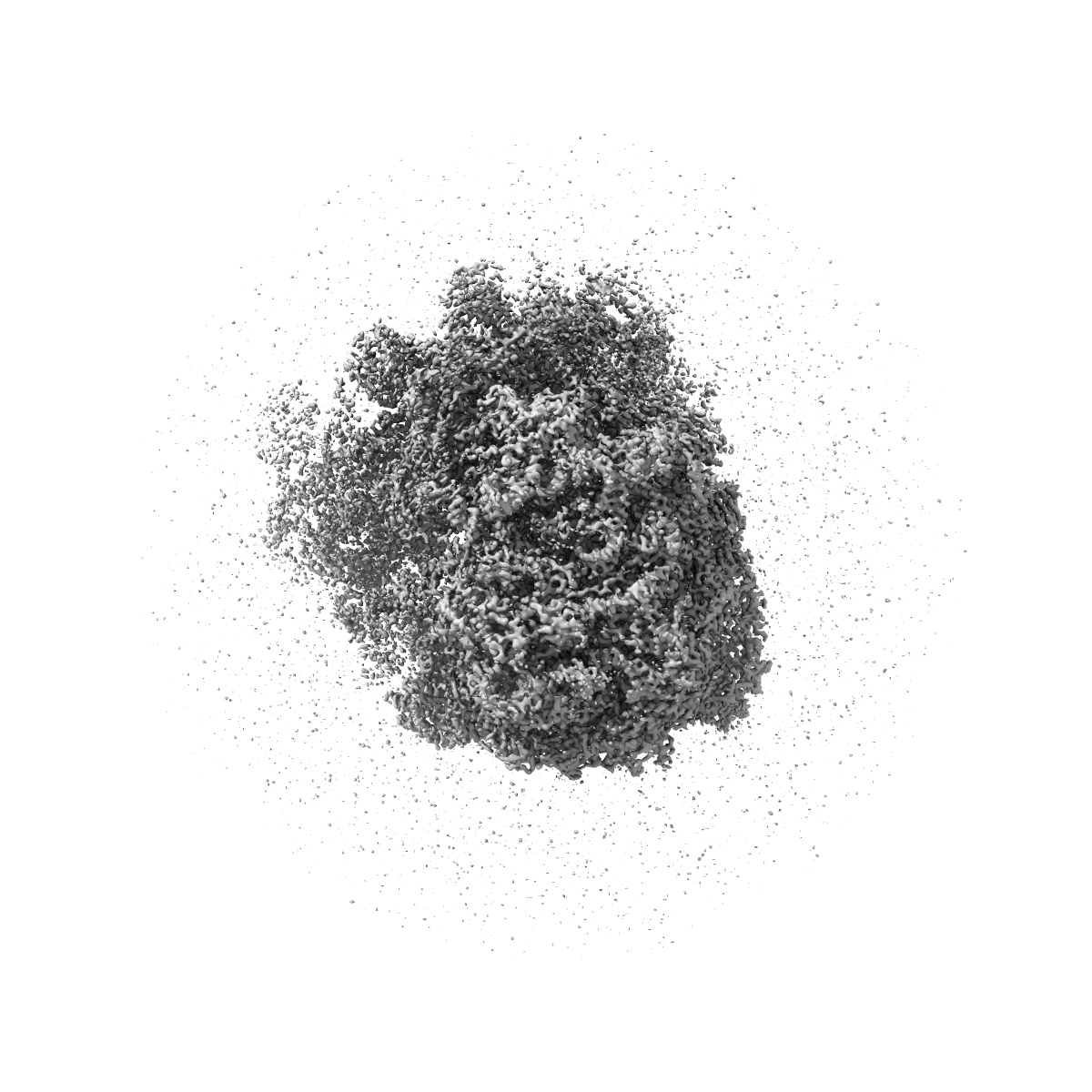
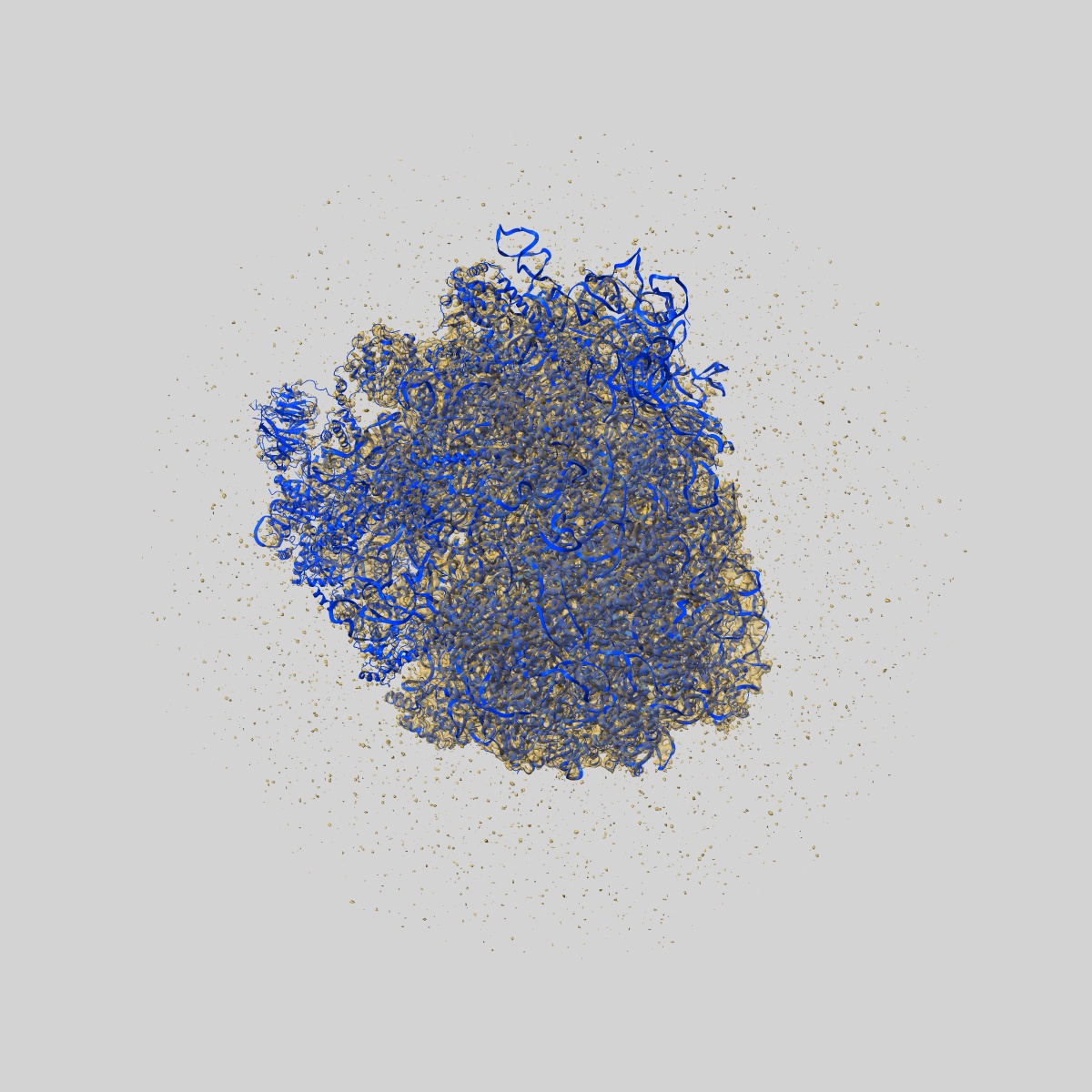
EMD-20952
K.lactis 80S ribosome with p/PE tRNA and eIF5B
EMD-20952
Single-particle3.6 Å
 Deposition: 14/11/2019
Map released: 15/01/2020
Last modified: 06/03/2024
Buffer
pH: 6.0
Grid
Model:
Quantifoil R2/2
Material: GOLD
Material: GOLD
Pretreatment
Support Film [1]
| Material | Topology | Thickness |
|---|---|---|
| CARBON | CONTINUOUS | 3.0 |
Vitrification
Microscope: FEI POLARA 300
Illumination mode: FLOOD BEAM
Imaging mode: BRIGHT FIELD
Electron source: FIELD EMISSION GUN
Acceleration voltage: 300 kV
Illumination mode: FLOOD BEAM
Imaging mode: BRIGHT FIELD
Electron source: FIELD EMISSION GUN
Acceleration voltage: 300 kV
Image Recording
[1]
Final
reconstruction
Resolution: 3.6
Å
(
BY AUTHOR)
Resolution method: FSC 0.143 CUT-OFF
Number of images used: 29712
Resolution method: FSC 0.143 CUT-OFF
Number of images used: 29712
⌯ Applied Symmetry
Point group:
C1
Startup model
[1]
Type:
EMDB MAP
⦨ Initial angle
assignment
Type:
ANGULAR RECONSTITUTION
⦩ Final angle assignment
Type:
ANGULAR RECONSTITUTION
Particle selection
[1]
| Selected | Ref. model | Method | Software | Details |
|---|---|---|---|---|
| 64815 | - | - | - | - |
Format: CCP4
Data type: IMAGE STORED AS FLOATING POINT NUMBER (4 BYTES)
Data type: IMAGE STORED AS FLOATING POINT NUMBER (4 BYTES)
⬡ Geometry
| X | Y | Z | |
|---|---|---|---|
| Origin | 0 | 0 | 0 |
| Dimensions (px) | 400 | 400 | 400 |
| Dimensions (Å) | 428.00003 | 428.00003 | 428.00003 |
| Voxel size (Å) | 1.07 | 1.07 | 1.07 |
Contour list
| Primary | Level | Source |
|---|---|---|
| True | 0.11 | AUTHOR |