EMD-2071
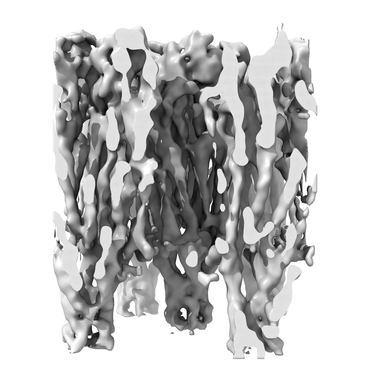
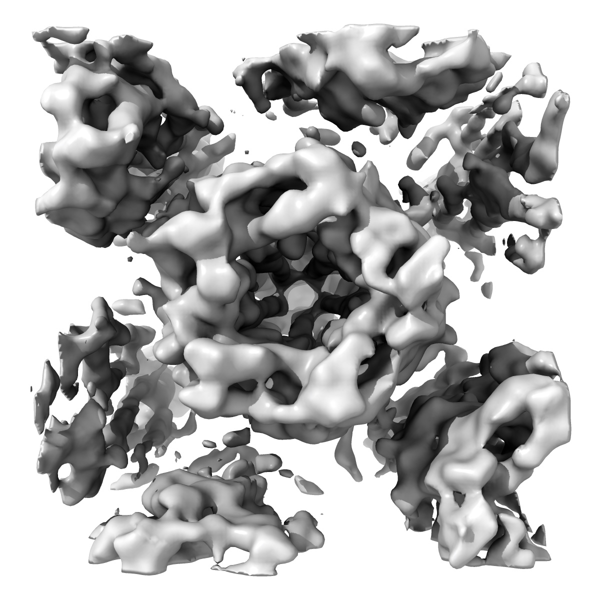
Gating movement in acetylcholine receptor analysed by time-resolved electron cryo-microscopy
EMD-2071
Helical reconstruction6.2 Å
 Deposition: 12/04/2012
Map released: 01/08/2012
Last modified: 26/09/2012
Details: Tubular membrane crystals of acetylcholine receptors grow spontaneously from isolated postsynaptic membranes when incubated in low salt buffer at 17 degrees C for two weeks
Buffer
pH: 7.0
Details: 100 mM sodium cacodylate, 1 mM calcium chloride
Details: 100 mM sodium cacodylate, 1 mM calcium chloride
Grid
Details: 300 mesh copper grid with pre-irradiated thick holey carbon support, glow discharged in amylamine atmosphere
Vitrification
Cryogen name: ETHANE
Chamber humidity: 85%
Chamber temperature: 120 K
Instrument: HOMEMADE PLUNGER
Method: Blot until applied droplet loses contact with filter paper (indicated by loss of transparency; typically 6s)
Timed resolved state: Vitrified within 10ms of exposure to acetylcholine (applied as the grid is being plunged,using a fine, focussed spray positioned about 1cm above the ethane surface)
Details: Vitrification carried out at an ambient temperature of 8 degrees C
Microscope: JEOL 3000SFF
Illumination mode: FLOOD BEAM
Imaging mode: BRIGHT FIELD
Electron source: FIELD EMISSION GUN
Acceleration voltage: 300 kV
Nominal CS: 1.6 mm
Nominal defocus: 0.9 µm - 2.0 µm
Nominal magnification: 40000.0
Calibrated magnification: 38500.0
Specimen holder model: OTHER
Specimen holder details: Top-entry holder for liquid helium cooled stage (the temperature of the specimen in this holder is usually at 4K)
Alignment procedure: LEGACY (Astigmatism: Objective lens astigmatism was corrected based on appearance of carbon film at 250,000 times magnification, Electron beam tilt params: )
Details: Standard low dose imaging of specimens over holes in the carbon support film
Temperature
Minimum: 10
K
Average: 10 K
Maximum: 20 K
Average: 10 K
Maximum: 20 K
Image Recording
[1]
Detector category:
FILM
Detector model: KODAK SO-163 FILM
Scanner: OTHER
Sampling interval: 2.5 µm
Number of real images: 111
Average electron dose per image: 25 e/Å2
Old range: 1.0
Bits per pixel: 16.0
Details: All images recorded on film, developed in Kodak d19 developer
Detector model: KODAK SO-163 FILM
Scanner: OTHER
Sampling interval: 2.5 µm
Number of real images: 111
Average electron dose per image: 25 e/Å2
Old range: 1.0
Bits per pixel: 16.0
Details: All images recorded on film, developed in Kodak d19 developer
Details: Alignment and distortion correction of each tube image was done using a segmental Fourier-Bessel method (Beroukhim & Unwin (1997) Ultramicroscopy, 70:57-81) with 50% overlap between successive segments
Final
reconstruction
Resolution: 6.2
Å
(
BY AUTHOR)
Resolution method: FSC 0.5 CUT-OFF
Algorithm: OTHER
Details: Final maps were calculated from 111 tube images(closed class) and 123 tube images (open class)
Resolution method: FSC 0.5 CUT-OFF
Algorithm: OTHER
Details: Final maps were calculated from 111 tube images(closed class) and 123 tube images (open class)
Software
[1]
| Name | Version | Details |
|---|---|---|
| MRC, and, own, programs | - | - |
CTF correction
Details: Each tube image
Format: CCP4
Data type: IMAGE STORED AS FLOATING POINT NUMBER (4 BYTES)
Annotation details: Density map of acetylcholine receptor
Details: ::::EMDATABANK.org::::EMD-2071::::
Data type: IMAGE STORED AS FLOATING POINT NUMBER (4 BYTES)
Annotation details: Density map of acetylcholine receptor
Details: ::::EMDATABANK.org::::EMD-2071::::
⬡ Geometry
| X | Y | Z | |
|---|---|---|---|
| Origin | 0 | 0 | 0 |
| Dimensions (px) | 128 | 128 | 168 |
| Dimensions (Å) | 128.0 | 128.0 | 168.0 |
| Voxel size (Å) | 1.0 | 1.0 | 1.0 |
Contour list
| Primary | Level | Source |
|---|---|---|
| True | 1.2 | AUTHOR |