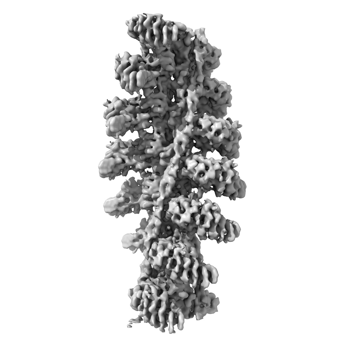
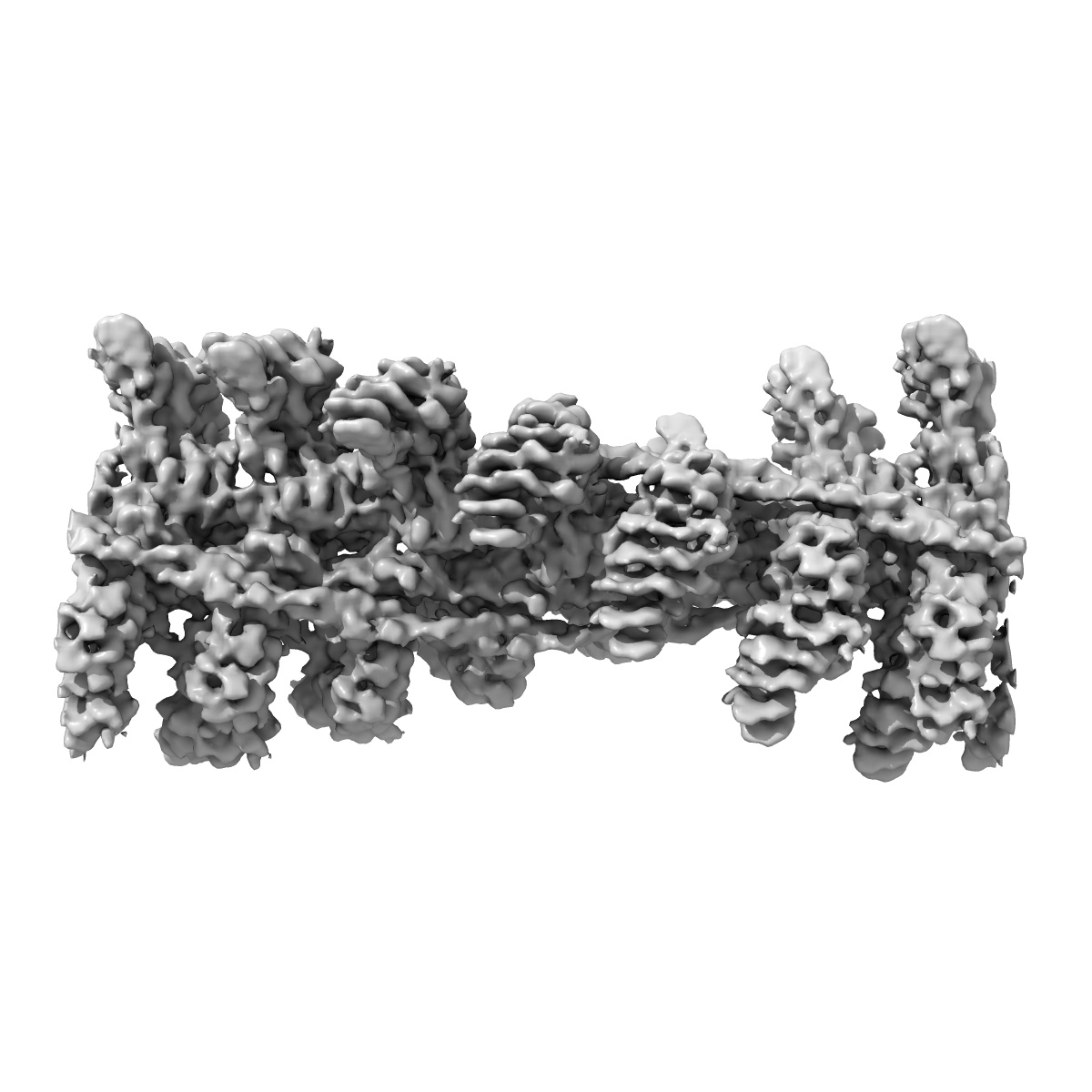
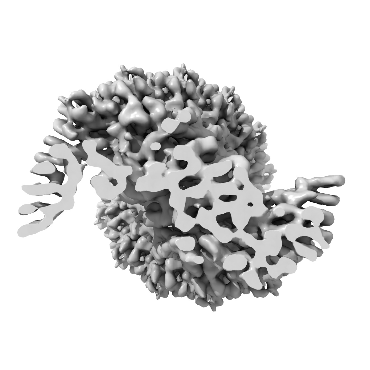
EMD-1987
Structure of the Actin-Tropomyosin-Myosin Complex (rigor ATM 3)
EMD-1987
Helical reconstruction7.7 Å
 Deposition: 14/11/2011
Map released: 01/08/2012
Last modified: 01/08/2012
Concentration: 0.01
mg/mL
Buffer
pH: 7.2
Details: 5mM Tris, 100mM KCl, 2mM MgCl2, 50mM glutamine, 50mM arginine
Details: 5mM Tris, 100mM KCl, 2mM MgCl2, 50mM glutamine, 50mM arginine
Grid
Details: C-Flat CF-2/1-4C copper 400 mesh
Vitrification
Cryogen name: ETHANE
Chamber humidity: 90%
Chamber temperature: 101 K
Instrument: GATAN CRYOPLUNGE 3
Method: Manual blotting for approximately 15 seconds
Details: Vitrification instrument: Gatan Cryoplunge 3
Chamber humidity: 90%
Chamber temperature: 101 K
Instrument: GATAN CRYOPLUNGE 3
Method: Manual blotting for approximately 15 seconds
Details: Vitrification instrument: Gatan Cryoplunge 3
Microscope: JEOL 3200FSC
Illumination mode: FLOOD BEAM
Imaging mode: BRIGHT FIELD
Electron source: FIELD EMISSION GUN
Acceleration voltage: 200 kV
Nominal CS: 4.1 mm
Nominal defocus: 0.75 µm - 1.5 µm
Nominal magnification: 80000.0
Calibrated magnification: 169644.0
Specimen holder model: JEOL 3200FSC CRYOHOLDER
Specimen holder details: Cryogenic stage with side entry access
Alignment procedure: LEGACY (Astigmatism: objective lens astigmatism was corrected at 150,000 times magnification, Electron beam tilt params: )
Temperature
Average: 77
K
Specialist optics
Energy filter
Image Recording
[1]
Detector category:
CCD
Detector model: TVIPS TEMCAM-F816 (8k x 8k)
Sampling interval: 15.6 µm
Number of real images: 836
Average electron dose per image: 17 e/Å2
Bits per pixel: 14.0
Details: Over 3000 images were taken of which only the best 836 were used for processing
Detector model: TVIPS TEMCAM-F816 (8k x 8k)
Sampling interval: 15.6 µm
Number of real images: 836
Average electron dose per image: 17 e/Å2
Bits per pixel: 14.0
Details: Over 3000 images were taken of which only the best 836 were used for processing
Details: Particles were selected by hand using e2helixboxer
Final
reconstruction
Resolution: 7.7
Å
(
BY AUTHOR)
Resolution method: FSC 0.5 CUT-OFF
Algorithm: OTHER
Details: Particles were classified using CODIM
Resolution method: FSC 0.5 CUT-OFF
Algorithm: OTHER
Details: Particles were classified using CODIM
⌯ Applied Symmetry
ΔΖ:
27.6
Å
ΔΦ: 166.5°
ΔΦ: 166.5°
Software
[1]
| Name | Version | Details |
|---|---|---|
| SPARX | - | - |
CTF correction
Details: Each particle
Format: CCP4
Data type: IMAGE STORED AS FLOATING POINT NUMBER (4 BYTES)
Annotation details: Conformation 3 of the F-actin-myo1E-tropomyosin complex
Details: ::::EMDATABANK.org::::EMD-1987::::
Data type: IMAGE STORED AS FLOATING POINT NUMBER (4 BYTES)
Annotation details: Conformation 3 of the F-actin-myo1E-tropomyosin complex
Details: ::::EMDATABANK.org::::EMD-1987::::
⬡ Geometry
| X | Y | Z | |
|---|---|---|---|
| Origin | 0 | 0 | 0 |
| Dimensions (px) | 160 | 160 | 220 |
| Dimensions (Å) | 294.4 | 294.4 | 404.80002 |
| Voxel size (Å) | 1.84 | 1.84 | 1.84 |
Contour list
| Primary | Level | Source |
|---|---|---|
| True | 0.0125 | AUTHOR |