EMD-15833

Helical reconstruction
2.58 Å
EMD-15833 Deposition: 16/09/2022
Map released: 24/05/2023
Last modified: 24/07/2024
Overview 3D View Sample Experiment Validation Additional data Links

EMD-15833

Archive Files (Depositor)

Primary 3D volume (map.gz) Half-map 1 Half-map 2 (map.gz) Experimental metadata (xml) Experimental metadata (cif.gz)

Validation (wwPDB)

Map-only validation report (pdf.gz) Map-8b3q summary report (pdf.gz) Map-8b3q FULL report (pdf.gz)

EMDB Annotations

EMICSS entry mapping (xml)

EMDB Files

VA raw map (map) VA resolution mask (mrc)

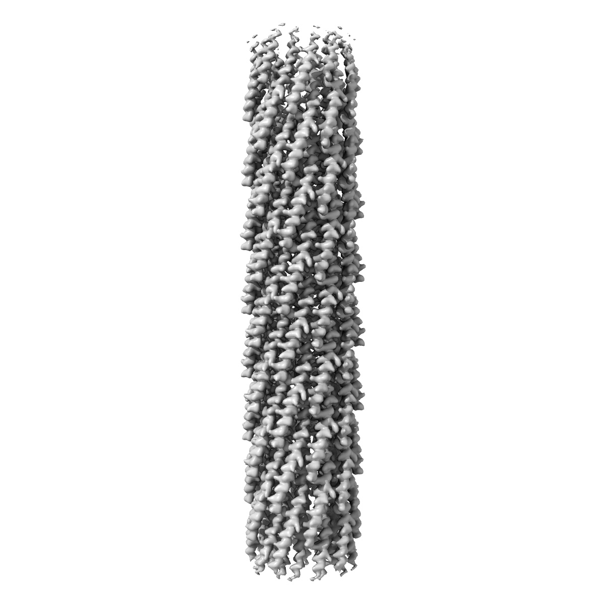
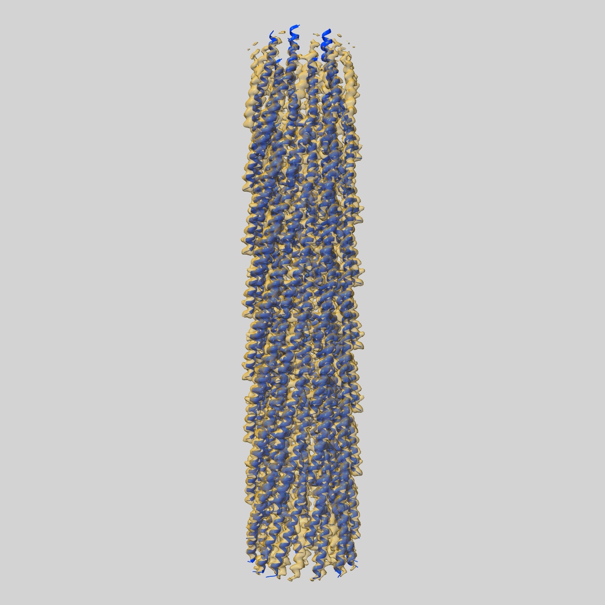
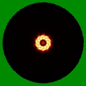
CryoEM structure of the central filamentous region of the f1 filamentous bacteriophage, consisting of the major capsid protein pVIII

EMD-15833

Helical reconstruction
2.58 Å
EMD-15833 Deposition: 16/09/2022
Map released: 24/05/2023
Last modified: 24/07/2024
Overview 3D View Sample Experiment Validation Additional data Links
Sample Organism: Enterobacteria phage f1
Sample: Enterobacteria phage f1
Fitted models: 8b3q

Deposition Authors: Conners R , McLaren M , Gold VAM
Cryo-electron microscopy of the f1 filamentous phage reveals insights into viral infection and assembly.
Conners R , Leon-Quezada RI , McLaren M , Bennett NJ , Daum B , Rakonjac J , Gold VAM
(2023) Nat Commun , 14 , 2724 - 2724
PUBMED: 37169795
DOI: doi:10.1038/s41467-023-37915-w
ISSN: 2041-1723
Grant Support: