



EMD-1527
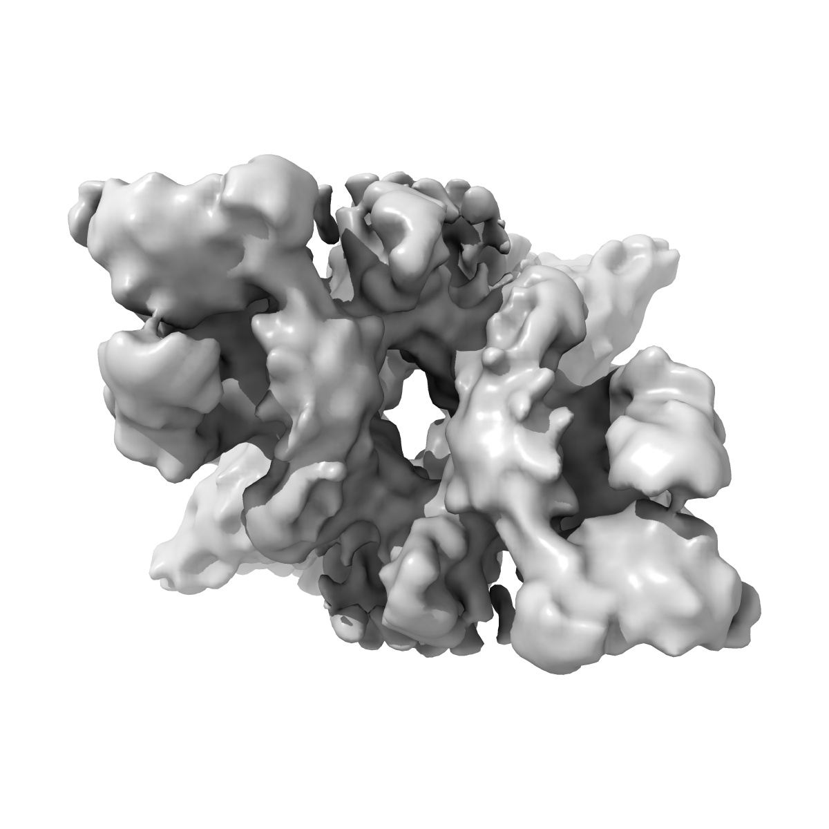
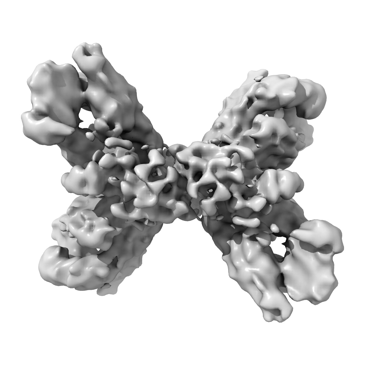
The structure of phosphorylase kinase holoenzyme at 9.9 A resolution and location of the catalytic subunit and the substrate glycogen phosphorylase
EMD-1527
Single-particle9.9 Å
 Deposition: 23/06/2008
Map released: 02/04/2009
Last modified: 24/10/2012
Concentration: 1.0
mg/mL
Buffer
pH: 8.2
Details: 100mM NaCl, 0.3 mM CaCl2, 5mM MgCl2, 50 mM Hepes, pH 8.2
Details: 100mM NaCl, 0.3 mM CaCl2, 5mM MgCl2, 50 mM Hepes, pH 8.2
Grid
Details: 400-mesh copper grid coated with a thin holey-carbon film
Vitrification
Cryogen name: ETHANE
Instrument: HOMEMADE PLUNGER
Method: Single-sided blotting
Details: Vitrification instrument: Manual. 5 microL were applied on a 200 mesh copper grid, coated with a thin holey carbon film. After blotting the excess of solution with Whatman paper, the grid was rapidly plunged into liquid ethane
Instrument: HOMEMADE PLUNGER
Method: Single-sided blotting
Details: Vitrification instrument: Manual. 5 microL were applied on a 200 mesh copper grid, coated with a thin holey carbon film. After blotting the excess of solution with Whatman paper, the grid was rapidly plunged into liquid ethane
Microscope: JEOL 2010UHR
Illumination mode: FLOOD BEAM
Imaging mode: BRIGHT FIELD
Electron source: FIELD EMISSION GUN
Acceleration voltage: 200 kV
Nominal CS: 0.5 mm
Nominal defocus: 2.8 µm - 4.8 µm
Nominal magnification: 50000.0
Specimen holder model: GATAN LIQUID NITROGEN
Specimen holder details: Gatan
Minimum tilt angle: 0
Maximum tilt angle: 0
Details: Low dose illumination
Illumination mode: FLOOD BEAM
Imaging mode: BRIGHT FIELD
Electron source: FIELD EMISSION GUN
Acceleration voltage: 200 kV
Nominal CS: 0.5 mm
Nominal defocus: 2.8 µm - 4.8 µm
Nominal magnification: 50000.0
Specimen holder model: GATAN LIQUID NITROGEN
Specimen holder details: Gatan
Minimum tilt angle: 0
Maximum tilt angle: 0
Details: Low dose illumination
Temperature
Minimum: 91.15
K
Average: 93.15 K
Maximum: 93.15 K
Average: 93.15 K
Maximum: 93.15 K
Image Recording
[1]
Detector category:
FILM
Detector model: KODAK SO-163 FILM
Scanner: NIKON SUPER COOLSCAN 9000
Sampling interval: 2.07 µm
Number of real images: 98
Average electron dose per image: 10 e/Å2
Bits per pixel: 8.0
Detector model: KODAK SO-163 FILM
Scanner: NIKON SUPER COOLSCAN 9000
Sampling interval: 2.07 µm
Number of real images: 98
Average electron dose per image: 10 e/Å2
Bits per pixel: 8.0
Details: The particles were selected using an automatic selection program
Final
reconstruction
Resolution: 9.9
Å
(
BY AUTHOR)
Resolution method: FSC 0.5 CUT-OFF
Number of images used: 18123
Algorithm: OTHER
Details: Final map was calculated using five groups of defocus
Resolution method: FSC 0.5 CUT-OFF
Number of images used: 18123
Algorithm: OTHER
Details: Final map was calculated using five groups of defocus
⌯ Applied Symmetry
Point group:
D2
Software
[1]
| Name | Version | Details |
|---|---|---|
| SPIDER | - | - |
CTF correction
Details: Wiener filtration of volumes from focal series
Format: CCP4
Data type: IMAGE STORED AS FLOATING POINT NUMBER (4 BYTES)
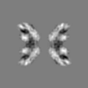
Annotation details: 3D map of PhK
Details: ::::EMDATABANK.org::::EMD-1527::::
Data type: IMAGE STORED AS FLOATING POINT NUMBER (4 BYTES)
Annotation details: 3D map of PhK
Details: ::::EMDATABANK.org::::EMD-1527::::
⬡ Geometry
| X | Y | Z | |
|---|---|---|---|
| Origin | 0 | 0 | 0 |
| Dimensions (px) | 180 | 180 | 180 |
| Dimensions (Å) | 372.6 | 372.6 | 372.6 |
| Voxel size (Å) | 2.07 | 2.07 | 2.07 |
Contour list
| Primary | Level | Source |
|---|---|---|
| True | 0.000757 | - |