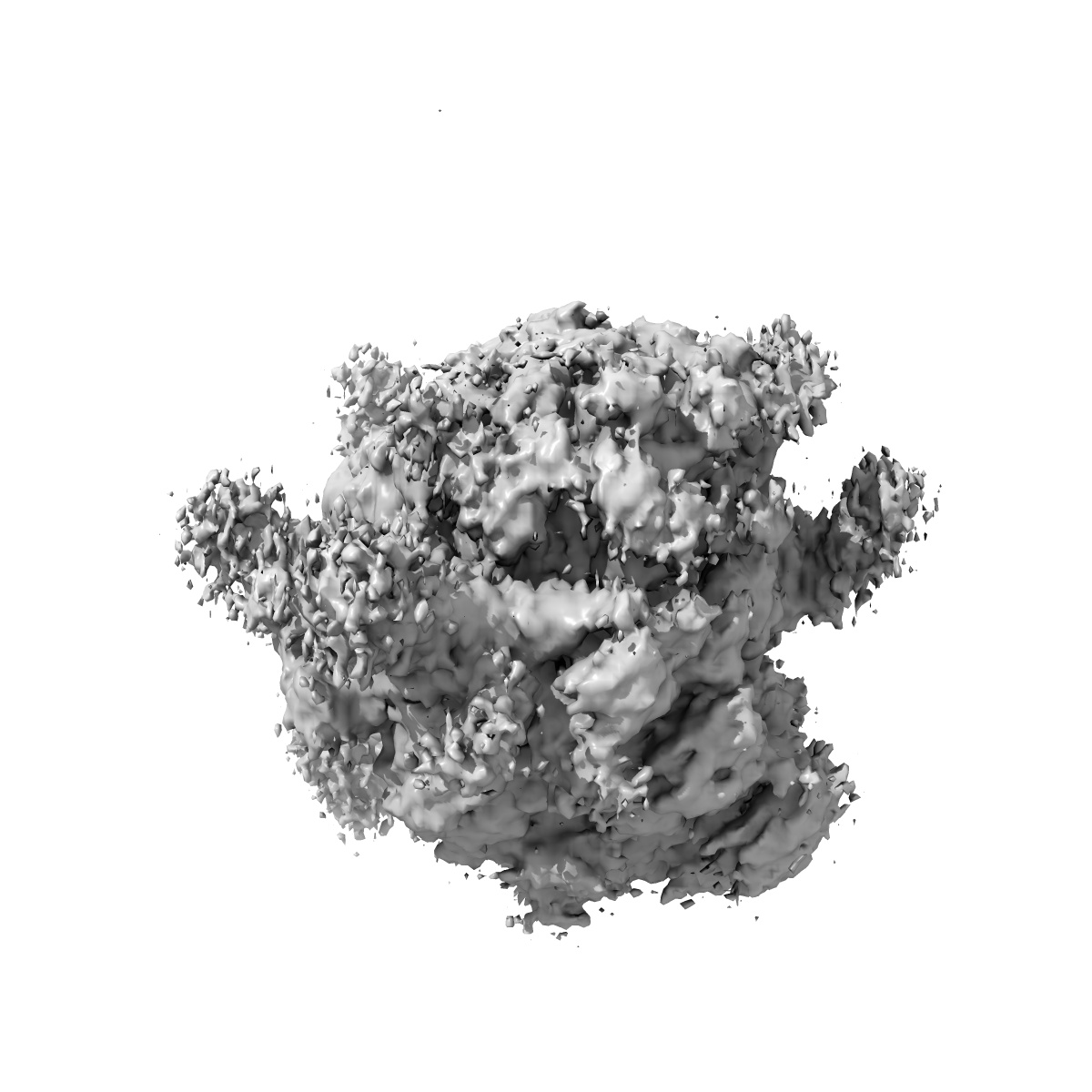
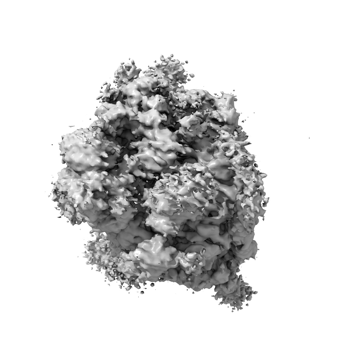
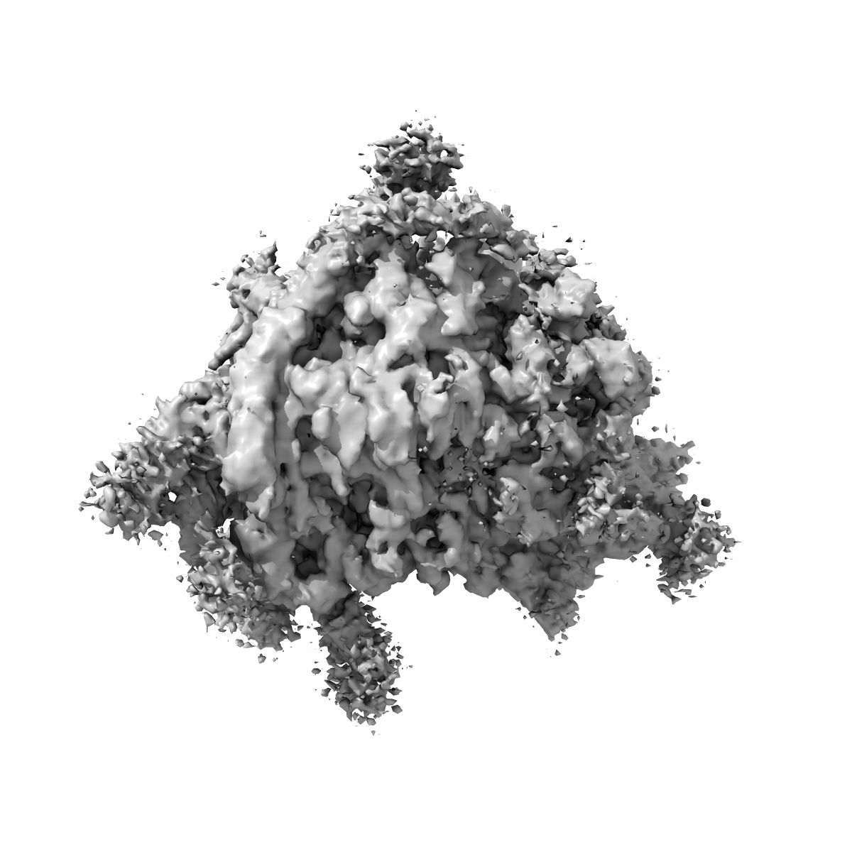
EMD-1480
3D structure of the canine 80S ribosome
EMD-1480
Single-particle8.7 Å
 Deposition: 24/02/2008
Map released: 31/03/2009
Last modified: 24/10/2012
Buffer
pH: 7.5
Details: 30mM Hepesm 50mM KAc, 10mM Mg acetate and 1.5% digitonin.
Details: 30mM Hepesm 50mM KAc, 10mM Mg acetate and 1.5% digitonin.
Grid
Details: 400
Vitrification
Cryogen name: ETHANE
Chamber humidity: 90%
Instrument: HOMEMADE PLUNGER
Method: blot for 1 second
Details: Vitrification instrument: home made plunger. in cold room
Chamber humidity: 90%
Instrument: HOMEMADE PLUNGER
Method: blot for 1 second
Details: Vitrification instrument: home made plunger. in cold room
Microscope: FEI TECNAI 20
Illumination mode: FLOOD BEAM
Imaging mode: BRIGHT FIELD
Electron source: FIELD EMISSION GUN
Acceleration voltage: 200 kV
Nominal CS: 2 mm
Nominal defocus: 0.5 µm - 3.0 µm
Nominal magnification: 50000.0
Calibrated magnification: 51000.0
Specimen holder model: OTHER
Specimen holder details: Eucentric
Details: Data were collected on Oxford and a Gatan cryo-holders
Temperature
Average: 93
K
Image Recording
[1]
Detector category:
FILM
Detector model: KODAK SO-163 FILM
Scanner: OTHER
Sampling interval: 4.5 µm
Number of real images: 500
Average electron dose per image: 20 e/Å2
Old range: 1.0
Bits per pixel: 16.0
Details: Creoscitex Eversmart was used to scan negatives.
Detector model: KODAK SO-163 FILM
Scanner: OTHER
Sampling interval: 4.5 µm
Number of real images: 500
Average electron dose per image: 20 e/Å2
Old range: 1.0
Bits per pixel: 16.0
Details: Creoscitex Eversmart was used to scan negatives.
Details: Particles selected with Boxer
Final
reconstruction
Resolution: 8.7
Å
(
BY AUTHOR)
Resolution method: FSC 0.5 CUT-OFF
Number of images used: 78800
Algorithm: OTHER
Details: Sep option equals 3 and setsf were used in the final cycle. a combined structure factor was used to correct for amplitudes in EMAN using the low resolution region from the images and the mid-resolution region from a low angle X-ray diffraction pattern.
⌯ Applied Symmetry
Point group:
C1
Software
[1]
| Name | Version | Details |
|---|---|---|
| EMAN | - | - |
⦩ Final angle assignment
Details: 2 degree steps between classes
CTF correction
Details: By micrograph
Format: CCP4
Data type: IMAGE STORED AS FLOATING POINT NUMBER (4 BYTES)
Annotation details: 3D volume of the canine 80S ribosome determined at 8.7 A resolution (Fsc 0.5), within the context of a ribosome-channel complex from ER membranes.
Details: ::::EMDATABANK.org::::EMD-1480::::
Data type: IMAGE STORED AS FLOATING POINT NUMBER (4 BYTES)
Annotation details: 3D volume of the canine 80S ribosome determined at 8.7 A resolution (Fsc 0.5), within the context of a ribosome-channel complex from ER membranes.
Details: ::::EMDATABANK.org::::EMD-1480::::
⬡ Geometry
| X | Y | Z | |
|---|---|---|---|
| Origin | 0 | 0 | 0 |
| Dimensions (px) | 168 | 168 | 168 |
| Dimensions (Å) | 458.64 | 458.64 | 458.64 |
| Voxel size (Å) | 2.73 | 2.73 | 2.73 |
Contour list
| Primary | Level | Source |
|---|---|---|
| True | 1.0 | - |