EMD-1273
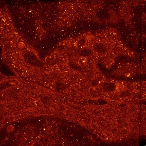
Centrosome polarization delivers secretory granules to the immunological synapse.
EMD-1273
Tomography50.0 Å
 Deposition: 09/10/2006
Map released: 10/10/2006
Last modified: 26/12/2012
| File | Description | Details |
|---|---|---|
| emd_1273_msk_5.map | Synaptic cleft, i.e. space between the CTL and the target cell as defined by the opposing membranes. Areas of close membrane-membrane contact (adhesion/signaling domains) and pocket-like areas with larger membrane-membrane distances (secretion domain) are clearly distinguishable | |
| emd_1273_msk_1.map | Microtubule (MT) network within the CTL. The MTs radiate out from the polarized centrosomal area, running both away from the plasma membrane into the cell and along the cell membrane to the periphery of the synapse. Both lytic granules (see mask granules_mask4) and the Golgi apparatus (see mask golgi_mask5) are linked to and transported along the MTs to the synaptic cleft | |
| emd_1273_msk_2.map | One of the two CTL centrioles, sitting close to the synaptic cleft. The two centrioles form part of the centrosome that is polarized to the synaptic cleft. Microtubules run from here into the cell and along the cell membrane to the periphery of the synapse (see mask MTs_mask) | |
| emd_1273_msk_3.map | The lytic granules are the "cytotoxic organelles" of the CTL. They are transported along the MTs in a minus-end fashion towards the polarized centrosome. After docking to the cell membrane they probably fuse with the membrane at the secretory domain and excrete the cytotoxic proteins into the synaptic cleft | |
| emd_1273_msk_4.map | A CTL Golgi apparatus, sitting close to the synaptic cleft. The Golgi apparatus is polarized to the cell-cell contact together with the centrosome |
×
Mask details
File: emd_1273_msk_5.map
Format: CCP4
Data type: IMAGE STORED AS SIGNED BYTE
Mask details: ::::EMDATABANK.org::::EMD-1273::::MASK:1::::
Format: CCP4
Data type: IMAGE STORED AS SIGNED BYTE
Mask details: ::::EMDATABANK.org::::EMD-1273::::MASK:1::::
⬡ Mask parameters
| X | Y | Z | |
|---|---|---|---|
| Dimensions | 2048 | 2048 | 76 |
| Origin | 0 | 0 | 0 |
| Spacing | 2048 | 2048 | 76 |
| Voxel size | 22.5 Å | 22.5 Å | 22.5 Å |
⌯ Symmetry
Space group: 1
⿴ Cell
A: 46080.0 Å
B: 46080.0 Å
C: 1710.0 Å
γ: 90.0°
α: 90.0°
β: 90.0°
B: 46080.0 Å
C: 1710.0 Å
γ: 90.0°
α: 90.0°
β: 90.0°
Map statistics
Minimum: 0.00e+00
Maximum: 1.00e+00
Average: 2.14e-02
Std: 1.45e-01
Maximum: 1.00e+00
Average: 2.14e-02
Std: 1.45e-01
×
Mask details
File: emd_1273_msk_1.map
Format: CCP4
Data type: IMAGE STORED AS SIGNED BYTE
Mask details: ::::EMDATABANK.org::::EMD-1273::::MASK:2::::
Format: CCP4
Data type: IMAGE STORED AS SIGNED BYTE
Mask details: ::::EMDATABANK.org::::EMD-1273::::MASK:2::::
⬡ Mask parameters
| X | Y | Z | |
|---|---|---|---|
| Dimensions | 2048 | 2048 | 76 |
| Origin | 0 | 0 | 0 |
| Spacing | 2048 | 2048 | 76 |
| Voxel size | 22.5 Å | 22.5 Å | 22.5 Å |
⌯ Symmetry
Space group: 1
⿴ Cell
A: 46080.0 Å
B: 46080.0 Å
C: 1710.0 Å
γ: 90.0°
α: 90.0°
β: 90.0°
B: 46080.0 Å
C: 1710.0 Å
γ: 90.0°
α: 90.0°
β: 90.0°
Map statistics
Minimum: 0.00e+00
Maximum: 1.00e+00
Average: 1.62e-03
Std: 4.02e-02
Maximum: 1.00e+00
Average: 1.62e-03
Std: 4.02e-02
×
Mask details
File: emd_1273_msk_2.map
Format: CCP4
Data type: IMAGE STORED AS SIGNED BYTE
Mask details: ::::EMDATABANK.org::::EMD-1273::::MASK:3::::
Format: CCP4
Data type: IMAGE STORED AS SIGNED BYTE
Mask details: ::::EMDATABANK.org::::EMD-1273::::MASK:3::::
⬡ Mask parameters
| X | Y | Z | |
|---|---|---|---|
| Dimensions | 2048 | 2048 | 76 |
| Origin | 0 | 0 | 0 |
| Spacing | 2048 | 2048 | 76 |
| Voxel size | 22.5 Å | 22.5 Å | 22.5 Å |
⌯ Symmetry
Space group: 1
⿴ Cell
A: 46080.0 Å
B: 46080.0 Å
C: 1710.0 Å
γ: 90.0°
α: 90.0°
β: 90.0°
B: 46080.0 Å
C: 1710.0 Å
γ: 90.0°
α: 90.0°
β: 90.0°
Map statistics
Minimum: 0.00e+00
Maximum: 1.00e+00
Average: 2.42e-04
Std: 1.56e-02
Maximum: 1.00e+00
Average: 2.42e-04
Std: 1.56e-02
×
Mask details
File: emd_1273_msk_3.map
Format: CCP4
Data type: IMAGE STORED AS SIGNED BYTE
Mask details: ::::EMDATABANK.org::::EMD-1273::::MASK:4::::
Format: CCP4
Data type: IMAGE STORED AS SIGNED BYTE
Mask details: ::::EMDATABANK.org::::EMD-1273::::MASK:4::::
⬡ Mask parameters
| X | Y | Z | |
|---|---|---|---|
| Dimensions | 2048 | 2048 | 76 |
| Origin | 0 | 0 | 0 |
| Spacing | 2048 | 2048 | 76 |
| Voxel size | 22.5 Å | 22.5 Å | 22.5 Å |
⌯ Symmetry
Space group: 1
⿴ Cell
A: 46080.0 Å
B: 46080.0 Å
C: 1710.0 Å
γ: 90.0°
α: 90.0°
β: 90.0°
B: 46080.0 Å
C: 1710.0 Å
γ: 90.0°
α: 90.0°
β: 90.0°
Map statistics
Minimum: 0.00e+00
Maximum: 1.00e+00
Average: 3.38e-02
Std: 1.81e-01
Maximum: 1.00e+00
Average: 3.38e-02
Std: 1.81e-01
×
Mask details
File: emd_1273_msk_4.map
Format: CCP4
Data type: IMAGE STORED AS SIGNED BYTE
Mask details: ::::EMDATABANK.org::::EMD-1273::::MASK:5::::
Format: CCP4
Data type: IMAGE STORED AS SIGNED BYTE
Mask details: ::::EMDATABANK.org::::EMD-1273::::MASK:5::::
⬡ Mask parameters
| X | Y | Z | |
|---|---|---|---|
| Dimensions | 2048 | 2048 | 76 |
| Origin | 0 | 0 | 0 |
| Spacing | 2048 | 2048 | 76 |
| Voxel size | 22.5 Å | 22.5 Å | 22.5 Å |
⌯ Symmetry
Space group: 1
⿴ Cell
A: 46080.0 Å
B: 46080.0 Å
C: 1710.0 Å
γ: 90.0°
α: 90.0°
β: 90.0°
B: 46080.0 Å
C: 1710.0 Å
γ: 90.0°
α: 90.0°
β: 90.0°
Map statistics
Minimum: 0.00e+00
Maximum: 1.00e+00
Average: 3.94e-03
Std: 6.26e-02
Maximum: 1.00e+00
Average: 3.94e-03
Std: 6.26e-02