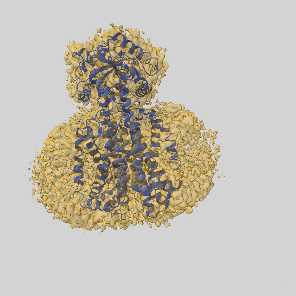
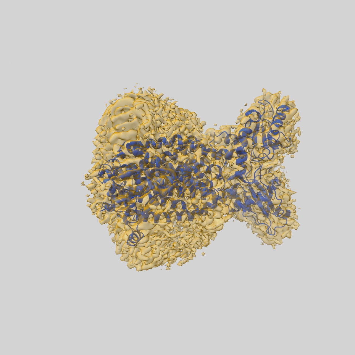
EMD-12604
Cryo-EM structure of the mycolic acid transporter MmpL3 from M. tuberculosis
EMD-12604
Single-particle3.0 Å
 Deposition: 15/03/2021
Map released: 16/06/2021
Last modified: 09/07/2025
Microscope: FEI TITAN KRIOS
Illumination mode: FLOOD BEAM
Imaging mode: BRIGHT FIELD
Electron source: FIELD EMISSION GUN
Acceleration voltage: 300 kV
Illumination mode: FLOOD BEAM
Imaging mode: BRIGHT FIELD
Electron source: FIELD EMISSION GUN
Acceleration voltage: 300 kV
Image Recording
[1]
Final
reconstruction
Startup model
[1]
Type:
NONE
⦨ Initial angle
assignment
Type:
MAXIMUM LIKELIHOOD
⦩ Final angle assignment
Type:
MAXIMUM LIKELIHOOD
CTF correction
Format: CCP4
Data type: IMAGE STORED AS FLOATING POINT NUMBER (4 BYTES)
Annotation details: Sharpened map
Data type: IMAGE STORED AS FLOATING POINT NUMBER (4 BYTES)
Annotation details: Sharpened map
⬡ Geometry
| X | Y | Z | |
|---|---|---|---|
| Origin | 0 | 0 | 0 |
| Dimensions (px) | 320 | 320 | 320 |
| Dimensions (Å) | 266.24 | 266.24 | 266.24 |
| Voxel size (Å) | 0.83199996 | 0.83199996 | 0.83199996 |
Contour list
| Primary | Level | Source |
|---|---|---|
| True | 0.4 | AUTHOR |