EMD-1069

Single-particle
21.0 Å
EMD-1069 Deposition: 10/03/2004
Map released: 11/03/2004
Last modified: 24/10/2012
Overview 3D View Sample Experiment Validation Additional data Links

EMD-1069

Archive Files (Depositor)

Primary 3D volume (map.gz) Experimental metadata (xml) Experimental metadata (cif.gz)

Validation (wwPDB)

Map-only validation report (pdf.gz)

EMDB Annotations

EMICSS entry mapping (xml)

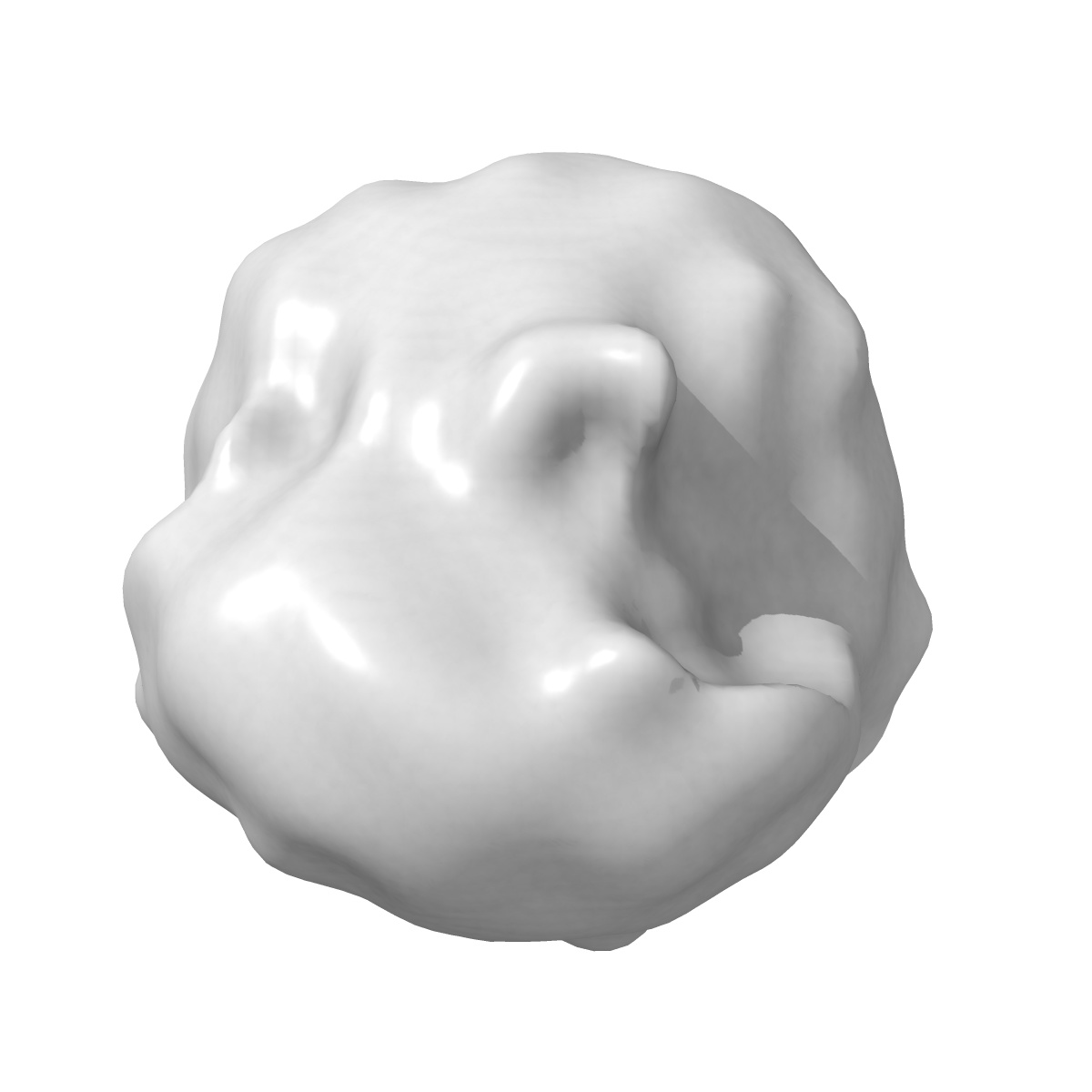
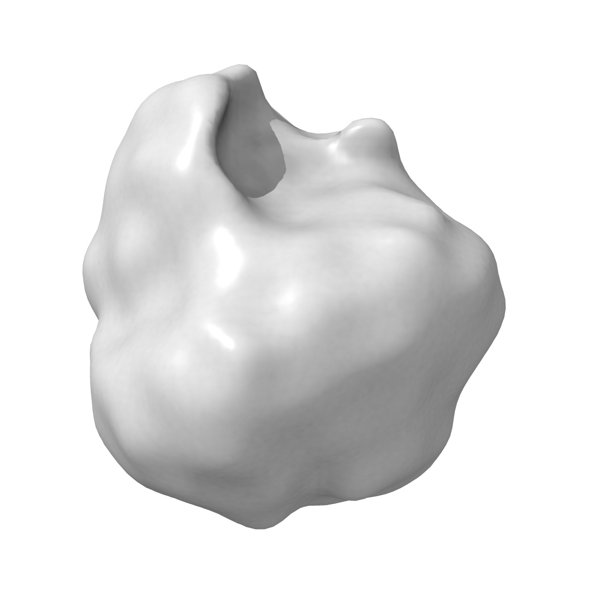
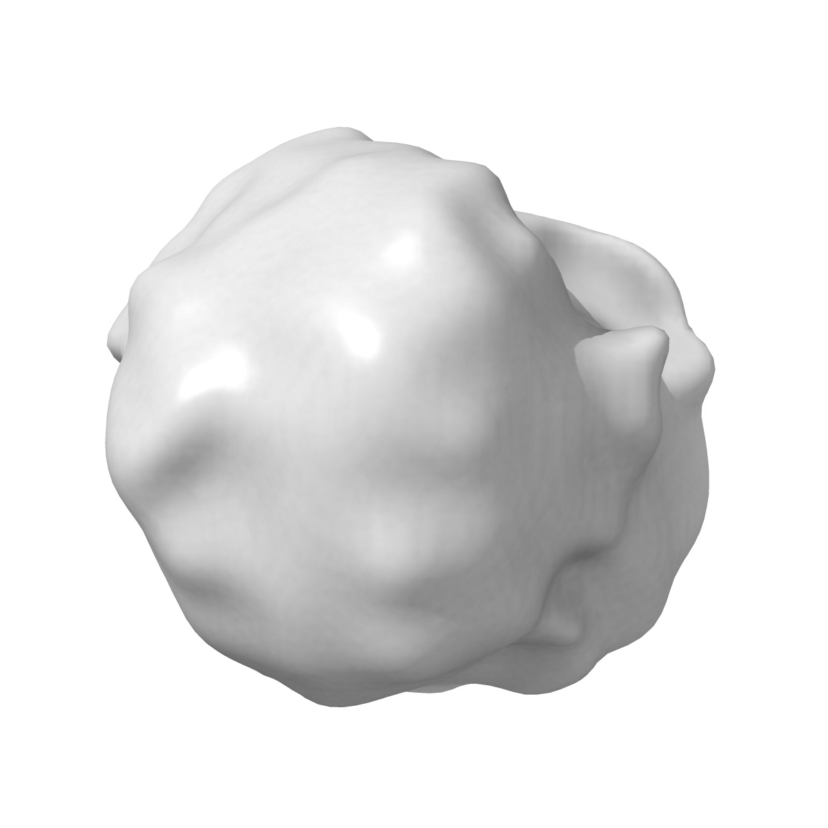
Visualization of the domain structure of an L-type Ca2+ channel using electron cryo-microscopy.

EMD-1069

Single-particle
21.0 Å
EMD-1069 Deposition: 10/03/2004
Map released: 11/03/2004
Last modified: 24/10/2012
Overview 3D View Sample Experiment Validation Additional data Links
Sample Organism: Oryctolagus cuniculus
Sample: skeletal muscle dihydropyridine receptor

Deposition Authors: Wolf M, Eberhart A, Glossmann H , Striessnig J , Grigorieff N
Visualization of the domain structure of an L-type Ca2+ channel using electron cryo-microscopy.
Wolf M, Eberhart A, Glossmann H , Striessnig J , Grigorieff N
(2003) J. Mol. Biol. , 332 , 171 - 182