EMD-10313

Single-particle
3.4 Å
EMD-10313 Deposition: 13/09/2019
Map released: 06/11/2019
Last modified: 22/05/2024
Overview 3D View Sample Experiment Validation Additional data Links

EMD-10313

Archive Files (Depositor)

Primary 3D volume (map.gz) Half-map 1 Half-map 2 (map.gz) Experimental metadata (xml) Experimental metadata (cif.gz)

Validation (wwPDB)

Map-only validation report (pdf.gz) Map-6suf summary report (pdf.gz) Map-6suf FULL report (pdf.gz)

EMDB Annotations

EMICSS entry mapping (xml)

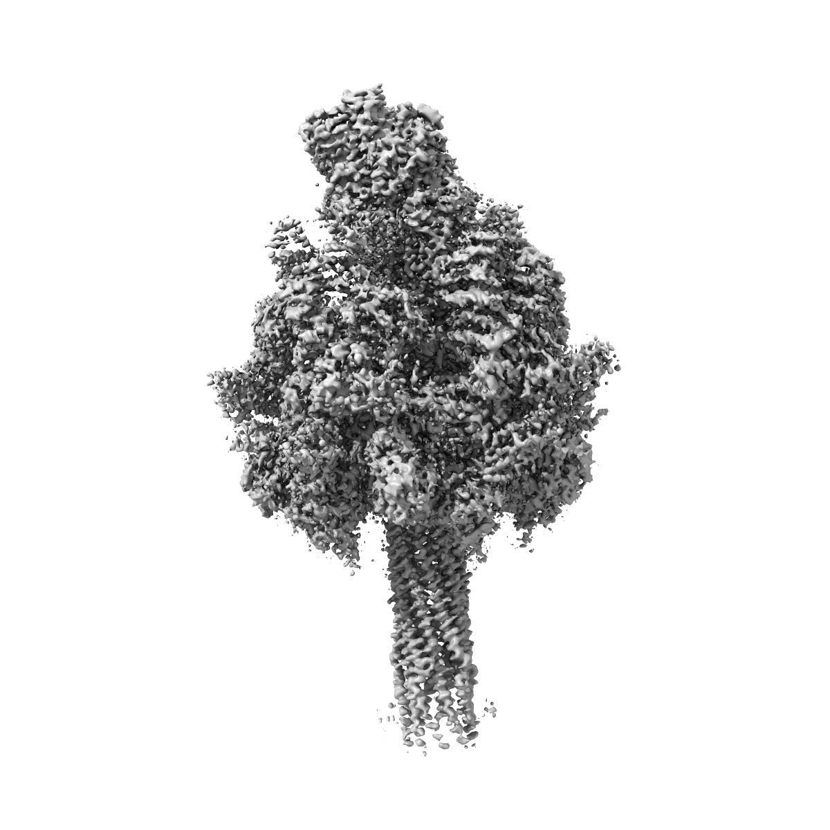
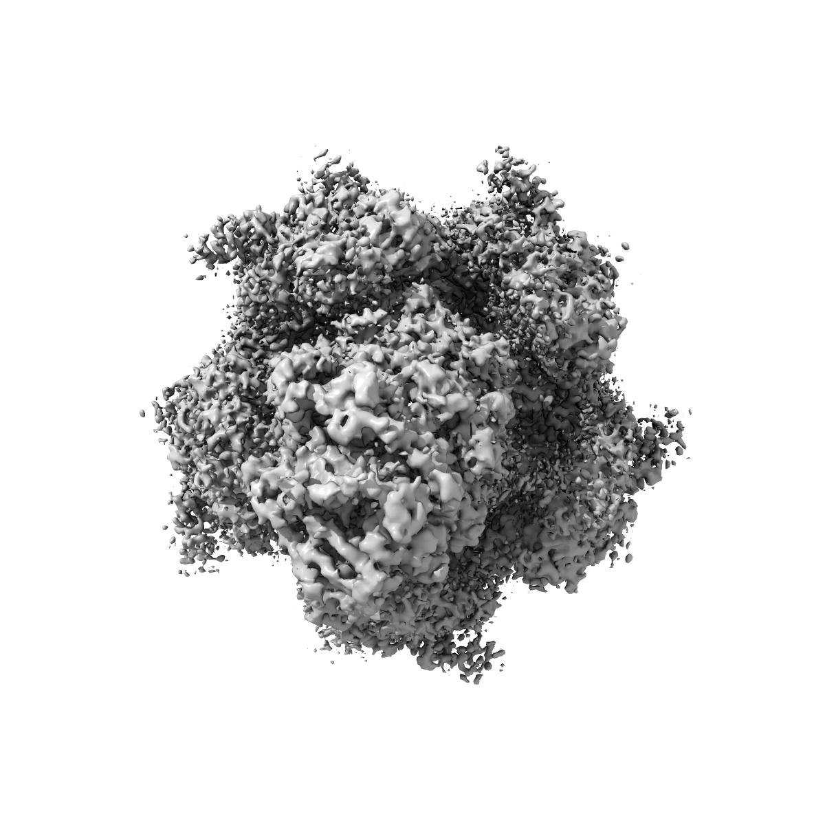
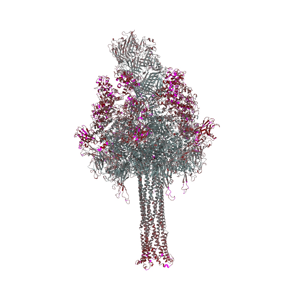
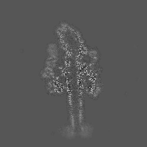
Structure of Photorhabdus luminescens Tc holotoxin pore

EMD-10313

Single-particle
3.4 Å
EMD-10313 Deposition: 13/09/2019
Map released: 06/11/2019
Last modified: 22/05/2024
Overview 3D View Sample Experiment Validation Additional data Links
Sample Organism: Photorhabdus luminescens
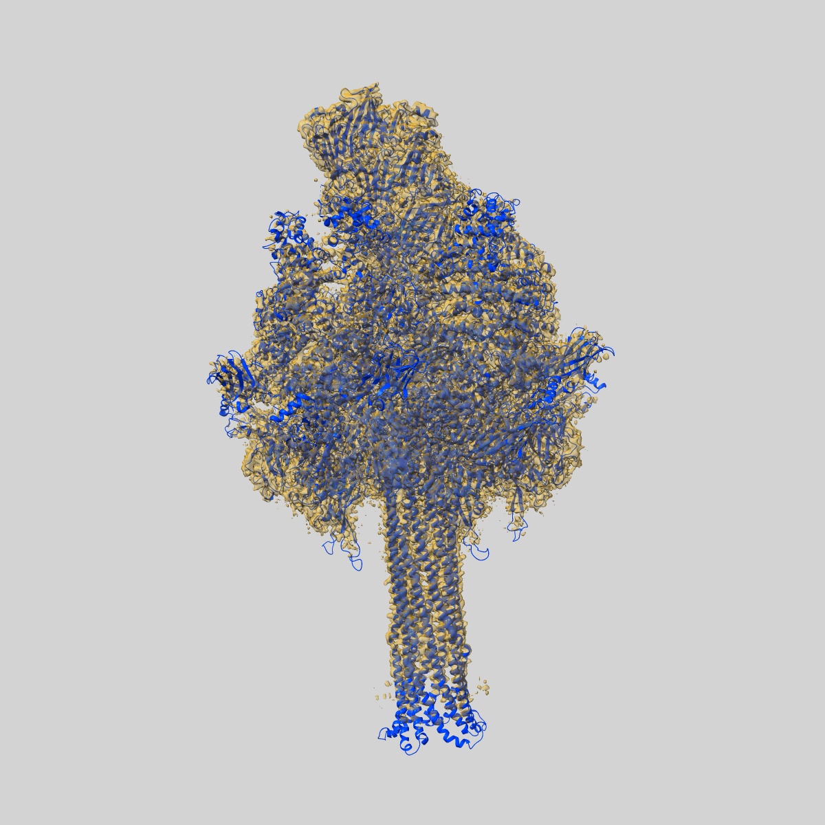
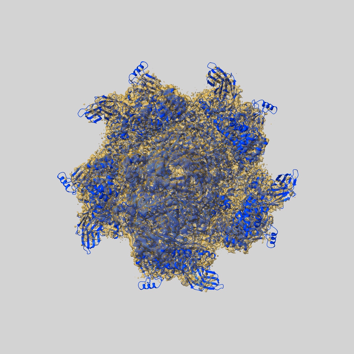
Sample: Tc holotoxin complex in the pore form, formed by the TcdA1 pentamer and TcdB2-TccC3
Fitted models: 6suf

Deposition Authors: Roderer D , Raunser S
Structure of a Tc holotoxin pore provides insights into the translocation mechanism.
Roderer D , Hofnagel O, Benz R, Raunser S
(2019) PNAS , 116 , 23083 - 23090
PUBMED: 31666324
DOI: doi:10.1073/pnas.1909821116
ISSN: 1091-6490
ASTM: PNASA6
Grant Support: