EMD-10069
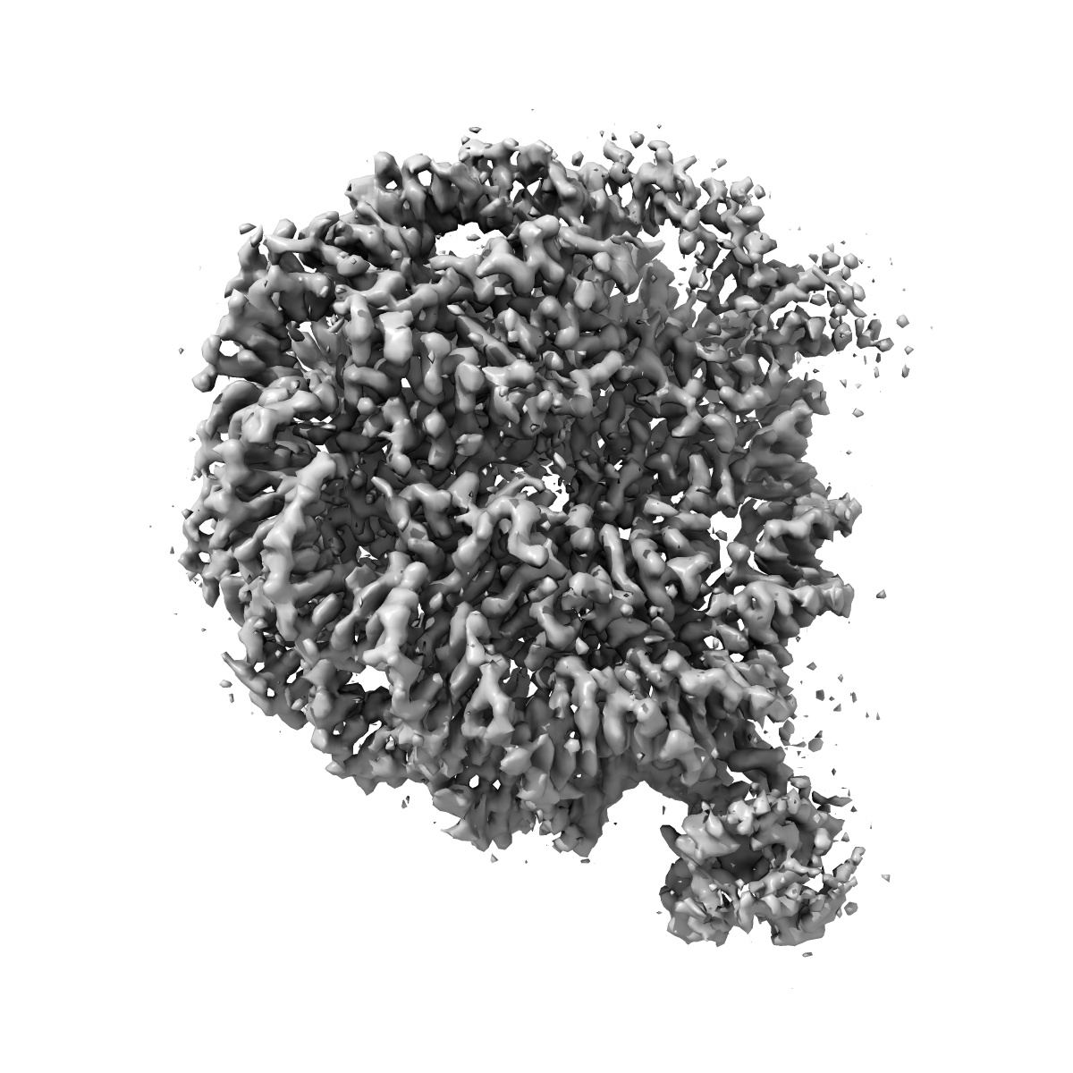
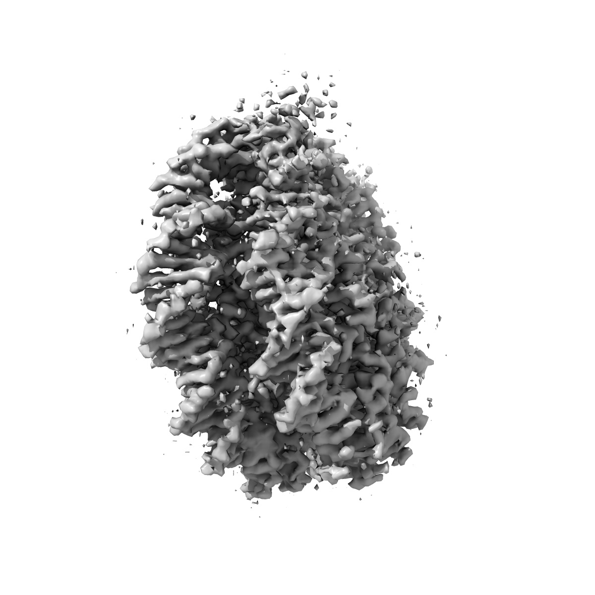
Structure of LEDGF PWWP domain bound H3K36 methylated nucleosome
EMD-10069
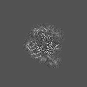
Single-particle3.2 Å
 Deposition: 13/06/2019
Map released: 18/12/2019
Last modified: 16/10/2024
Concentration: 0.2
mg/mL
Details: The complex is purified by gel filtration
Details: The complex is purified by gel filtration
Buffer
pH: 7.5
Buffer components [3]:
Details: Solution were made from stock solution
Buffer components [3]:
| Name | Formula | Concentration | ChEBI |
|---|---|---|---|
| HEPES | C8H18N2O4S | 20.0 mM | |
| Sodium chloride | NaCl | 50.0 mM | |
| Dithiothreitol | C4H10O2S2 | 1.0 mM |
Grid
Vitrification
Cryogen name: ETHANE
Chamber humidity: 100%
Chamber temperature: 277 K
Instrument: FEI VITROBOT MARK IV
Details: Blot for 4 seconds before plunging.
Chamber humidity: 100%
Chamber temperature: 277 K
Instrument: FEI VITROBOT MARK IV
Details: Blot for 4 seconds before plunging.
Microscope: FEI TITAN KRIOS
Illumination mode: FLOOD BEAM
Imaging mode: BRIGHT FIELD
Electron source: FIELD EMISSION GUN
Acceleration voltage: 300 kV
C2 aperture diameter: 70.0 µm
Nominal CS: 2.7 mm
Specimen holder model: FEI TITAN KRIOS AUTOGRID HOLDER
Alignment procedure: COMA FREE
Illumination mode: FLOOD BEAM
Imaging mode: BRIGHT FIELD
Electron source: FIELD EMISSION GUN
Acceleration voltage: 300 kV
C2 aperture diameter: 70.0 µm
Nominal CS: 2.7 mm
Specimen holder model: FEI TITAN KRIOS AUTOGRID HOLDER
Alignment procedure: COMA FREE
Image Recording
[1]
Detector model:
GATAN K2 SUMMIT (4k x 4k)
Detector mode: COUNTING
Average exposure time: 9.0 s
Average electron dose per image: 43.18 e/Å2
Detector mode: COUNTING
Average exposure time: 9.0 s
Average electron dose per image: 43.18 e/Å2
Final
reconstruction
Resolution: 3.2
Å
(
BY AUTHOR)
Resolution method: FSC 0.143 CUT-OFF
Number of classed used: 1
Number of images used: 55142
Resolution method: FSC 0.143 CUT-OFF
Number of classed used: 1
Number of images used: 55142
⌯ Applied Symmetry
Point group:
C1
Software
[1]
| Name | Version | Details |
|---|---|---|
| RELION | 3.0.5 | - |
Startup model
[1]
Type:
INSILICO MODEL
Insilico model: CryoSPARC ab-initio reconstruction was use to generate the startup model.
Insilico model: CryoSPARC ab-initio reconstruction was use to generate the startup model.
⦨ Initial angle
assignment
Type:
ANGULAR RECONSTITUTION
⦩ Final angle assignment
Particle selection
[1]
| Selected | Ref. model | Method | Software | Details |
|---|---|---|---|---|
| 527640 | - | - | - | - |
Format: CCP4
Data type: IMAGE STORED AS FLOATING POINT NUMBER (4 BYTES)
Annotation details: Cryo-EM structure of LEDGF bound H3K36me3 modified nucleosome.
Data type: IMAGE STORED AS FLOATING POINT NUMBER (4 BYTES)
Annotation details: Cryo-EM structure of LEDGF bound H3K36me3 modified nucleosome.
⬡ Geometry
| X | Y | Z | |
|---|---|---|---|
| Origin | 0 | 0 | 0 |
| Dimensions (px) | 256 | 256 | 256 |
| Dimensions (Å) | 268.8 | 268.8 | 268.8 |
| Voxel size (Å) | 1.05 | 1.05 | 1.05 |
Contour list
| Primary | Level | Source |
|---|---|---|
| True | 0.05 | AUTHOR |