EMD-0553
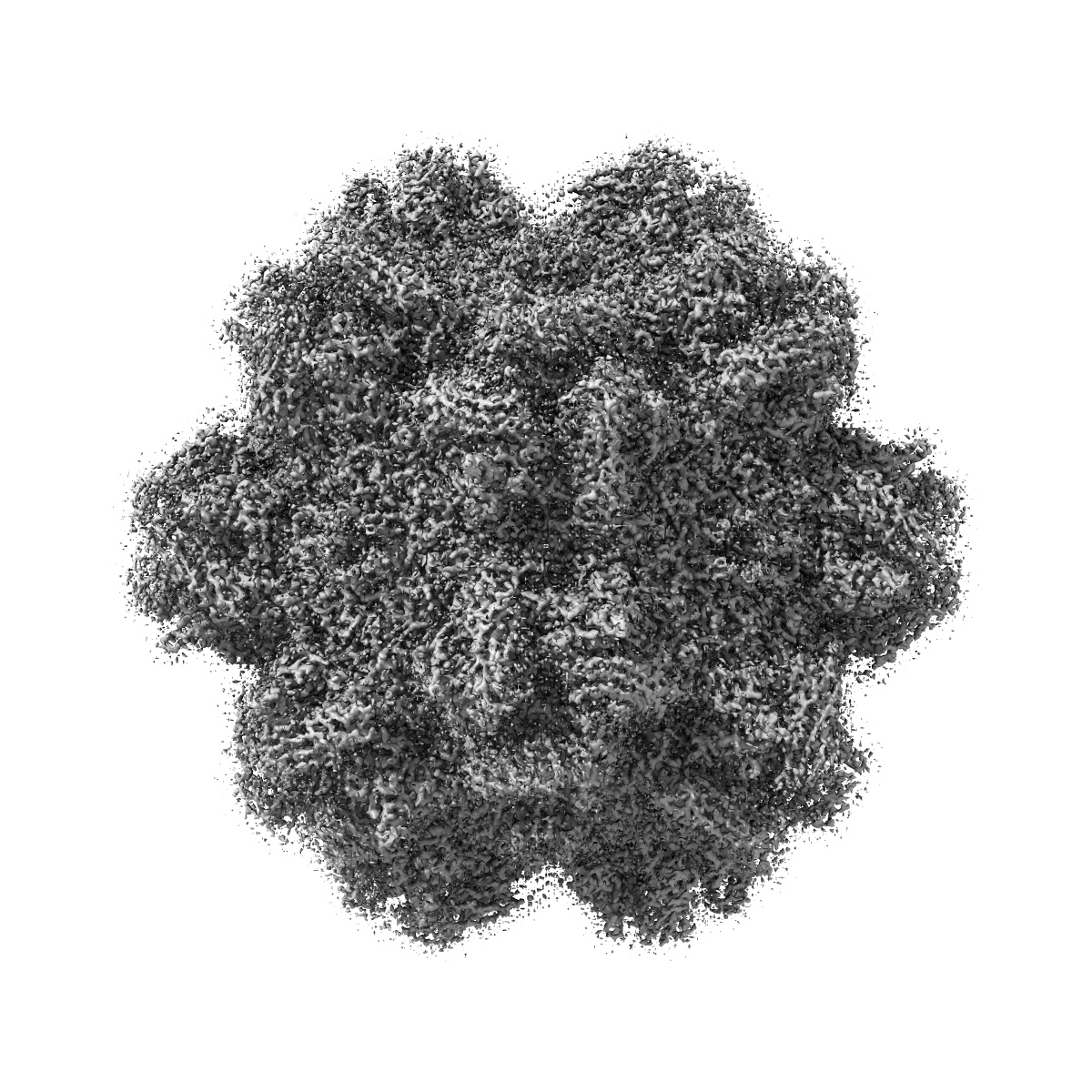
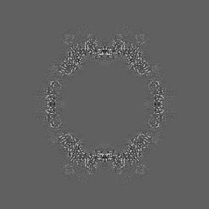
Cryo-EM structure of AAV-2 in complex with AAVR PKD domains 1 and 2
EMD-0553
Single-particle2.4 Å
 Deposition: 12/02/2019
Map released: 12/06/2019
Last modified: 16/10/2024
Concentration: 0.1
mg/mL
Details: Virus-like particles at 0.10 mg/mL were adhered to thin continuous carbon-coated grids, then, after initial blotting, AAVR-PKD1-2 was added at 0.74 mg/mL (a 10-fold molar excess over AAV2 subunits) before blotting again.
Details: Virus-like particles at 0.10 mg/mL were adhered to thin continuous carbon-coated grids, then, after initial blotting, AAVR-PKD1-2 was added at 0.74 mg/mL (a 10-fold molar excess over AAV2 subunits) before blotting again.
Buffer
Grid
Mesh: 400
Material: COPPER
Details: Glow discharge settings: Pelco easiGlow, 25s, 25mA, 0.39mBar, negative polarity Grid: Ted Pella Cat#01824, ultrathin carbon film (<3nm) on lacey carbon support film
Material: COPPER
Details: Glow discharge settings: Pelco easiGlow, 25s, 25mA, 0.39mBar, negative polarity Grid: Ted Pella Cat#01824, ultrathin carbon film (<3nm) on lacey carbon support film
Pretreatment
Vitrification
Cryogen name: ETHANE
Chamber humidity: 100%
Chamber temperature: 298.15 K
Instrument: FEI VITROBOT MARK IV
Details: 4ul of AAV-2 (1.7uM) was added to the grid, followed by 4ul of AAVR-PKD1-2 (16.7uM), with manual blotting after each addition with Whatman paper (Cat. no. 1001-110.) 4ul of buffer containing 25mM HEPES, 150mM NaCl, pH 7.4 was added to the grid before final blotting and plunge-freezing in the Vitrobot (blot time 1.5sec, blot force -1)..
Microscope: FEI TITAN KRIOS
Illumination mode: FLOOD BEAM
Imaging mode: BRIGHT FIELD
Electron source: FIELD EMISSION GUN
Acceleration voltage: 300 kV
Nominal CS: 2.7 mm
Nominal defocus: -0.8 µm - -2.0 µm
Nominal magnification: 75000.0
Specimen holder model: FEI TITAN KRIOS AUTOGRID HOLDER
Cooling holder cryogen: NITROGEN
Alignment procedure: COMA FREE
Image Recording
[1]
Detector model:
FEI FALCON III (4k x 4k)
Detector mode: SUPER-RESOLUTION
Dimensions: 7676 pixel x 7420 pixel
Number of grids: 1
Number of real images: 2329
Average exposure time: 40.0 s
Average electron dose per image: 25.4 e/Å2
Detector mode: SUPER-RESOLUTION
Dimensions: 7676 pixel x 7420 pixel
Number of grids: 1
Number of real images: 2329
Average exposure time: 40.0 s
Average electron dose per image: 25.4 e/Å2
Final
reconstruction
Resolution: 2.4
Å
(
BY AUTHOR)
Resolution method: FSC 0.143 CUT-OFF
Number of classed used: 1
Number of images used: 21373
Resolution method: FSC 0.143 CUT-OFF
Number of classed used: 1
Number of images used: 21373
⌯ Applied Symmetry
Point group:
I
Software
[1]
| Name | Version | Details |
|---|---|---|
| RELION | 3.0b | - |
Startup model
[1]
Type:
OTHER
⦨ Initial angle
assignment
⦩ Final angle assignment
Particle selection
[1]
| Selected | Ref. model | Method | Software | Details |
|---|---|---|---|---|
| 34450 | - | - | - | RELION Autopicker using 3D AAV-2 reference filtered to 20A with 15 degree sampling |
Final 3D classification
Number of classes:
2
Software
[1]
| Name | Version | Details |
|---|---|---|
| RELION | 3.0b | - |
Format: CCP4
Data type: IMAGE STORED AS FLOATING POINT NUMBER (4 BYTES)
Annotation details: EM sharpened map
Data type: IMAGE STORED AS FLOATING POINT NUMBER (4 BYTES)
Annotation details: EM sharpened map
⬡ Geometry
| X | Y | Z | |
|---|---|---|---|
| Origin | -192 | -192 | -192 |
| Dimensions (px) | 384 | 384 | 384 |
| Dimensions (Å) | 401.664 | 401.664 | 401.664 |
| Voxel size (Å) | 1.046 | 1.046 | 1.046 |
Contour list
| Primary | Level | Source |
|---|---|---|
| True | 0.053 | AUTHOR |