EMD-0160
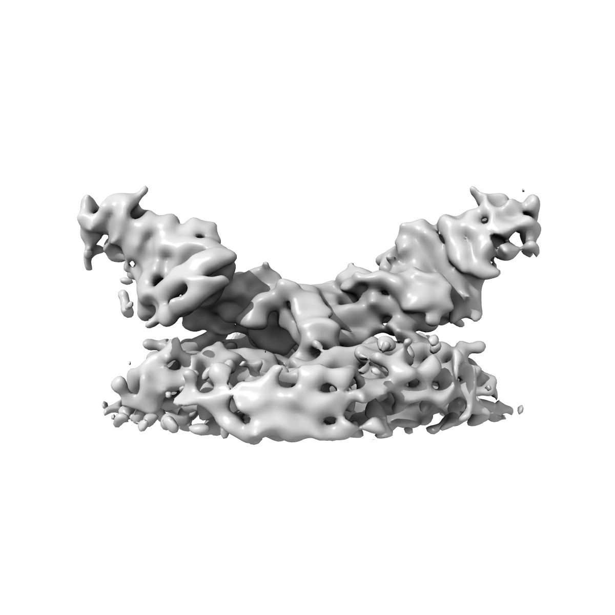
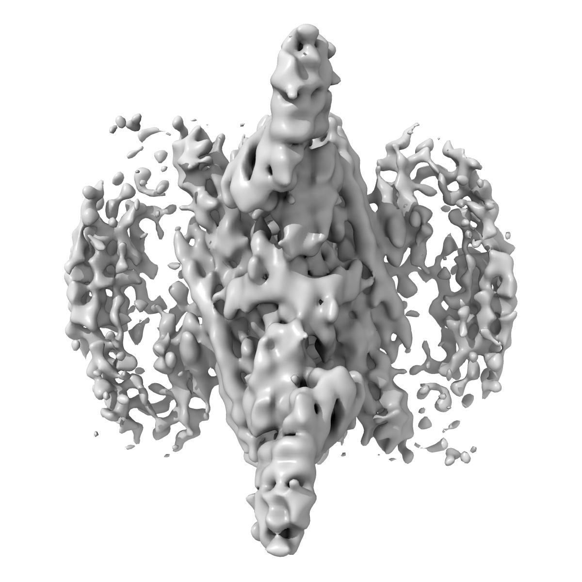
Retromer-Vps5: map centred on the Vps26 dimer.
EMD-0160
Subtomogram averaging8.9 Å
 Deposition: 31/07/2018
Map released: 26/09/2018
Last modified: 10/10/2018
Concentration: 1.1
mg/mL
Details: The solution-assembled complex was incubated with Folch liposomes at room temperature.
Details: The solution-assembled complex was incubated with Folch liposomes at room temperature.
Buffer
pH: 7.5
Buffer components [2]:
Buffer components [2]:
| Name | Formula | Concentration | ChEBI |
|---|---|---|---|
| HEPES-KOH | C8H19KN2O5S | 20.0 mM | |
| Sodium chloride | NaCl | 200.0 mM |
Vitrification
Microscope: FEI TITAN KRIOS
Illumination mode: FLOOD BEAM
Imaging mode: BRIGHT FIELD
Electron source: FIELD EMISSION GUN
Acceleration voltage: 300 kV
C2 aperture diameter: 70.0 µm
Nominal CS: 2.7 mm
Nominal defocus: 2.5 µm - 6.5 µm
Nominal magnification: 105000.0
Specimen holder model: FEI TITAN KRIOS AUTOGRID HOLDER
Cooling holder cryogen: NITROGEN
Alignment procedure: ZEMLIN TABLEAU
Specialist optics
Energy filter
Image Recording
[1]
Detector model:
GATAN K2 QUANTUM (4k x 4k)
Detector mode: SUPER-RESOLUTION
Number of grids: 2
Average electron dose per image: 3.17 e/Å2
Details: Tomographic tilt series were acquired with the dose-symmetric tilt-scheme (Hagen et al., J Struct Biol. 2017)
Detector mode: SUPER-RESOLUTION
Number of grids: 2
Average electron dose per image: 3.17 e/Å2
Details: Tomographic tilt series were acquired with the dose-symmetric tilt-scheme (Hagen et al., J Struct Biol. 2017)
Details: After motion correction in "alignframes" (IMOD), each of the images in the tilt series was low-pass filtered according to the electron-dose acquired by the sample (Grant and Grigorieff, 2015).
Final
reconstruction
Resolution: 8.9
Å
(
BY AUTHOR)
Resolution method: FSC 0.143 CUT-OFF
Algorithm: BACK PROJECTION
Details:
Resolution method: FSC 0.143 CUT-OFF
Algorithm: BACK PROJECTION
Details:
⌯ Applied Symmetry
Point group:
C2
⦩ Final angle assignment
Extraction
Number of images used: 194885
Reference model: Reference-free
Method: Geometrical seeding alogn the surface of manually traced tubules
Reference model: Reference-free
Method: Geometrical seeding alogn the surface of manually traced tubules
Format: CCP4
Data type: IMAGE STORED AS FLOATING POINT NUMBER (4 BYTES)
Annotation details: Retromer-Vps5: map centred on the Vps26 dimer.
Data type: IMAGE STORED AS FLOATING POINT NUMBER (4 BYTES)
Annotation details: Retromer-Vps5: map centred on the Vps26 dimer.
⬡ Geometry
| X | Y | Z | |
|---|---|---|---|
| Origin | 0 | 0 | 0 |
| Dimensions (px) | 180 | 180 | 180 |
| Dimensions (Å) | 243.0 | 243.0 | 243.0 |
| Voxel size (Å) | 1.35 | 1.35 | 1.35 |
Contour list
| Primary | Level | Source |
|---|---|---|
| True | 0.15 | AUTHOR |