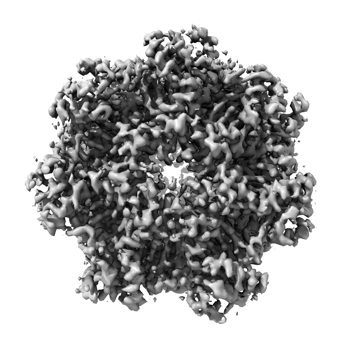
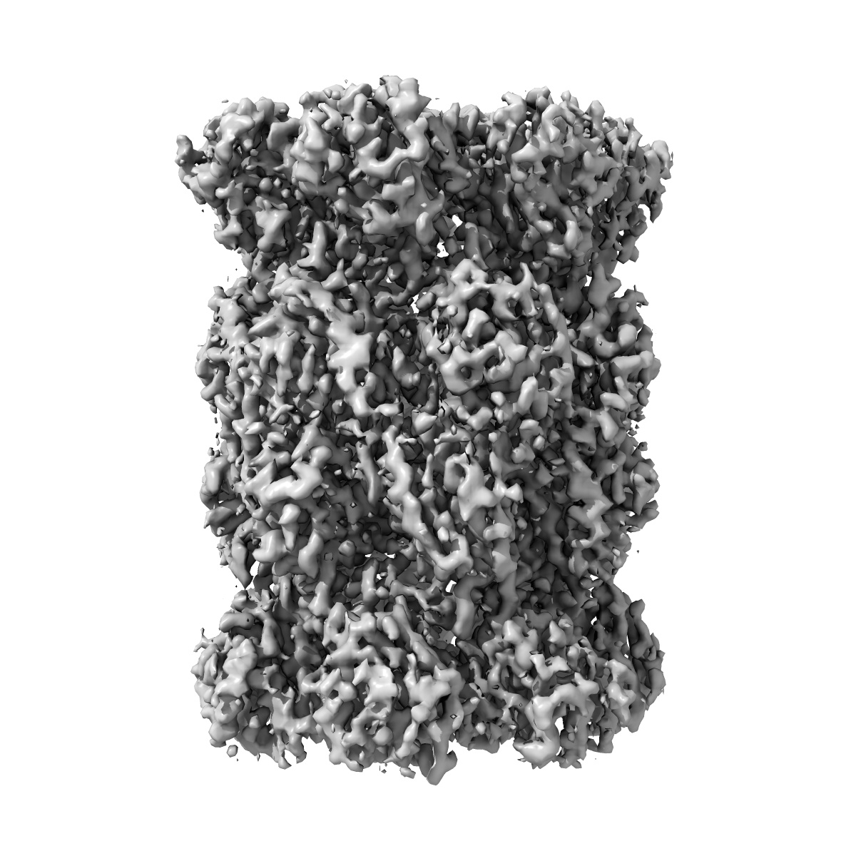
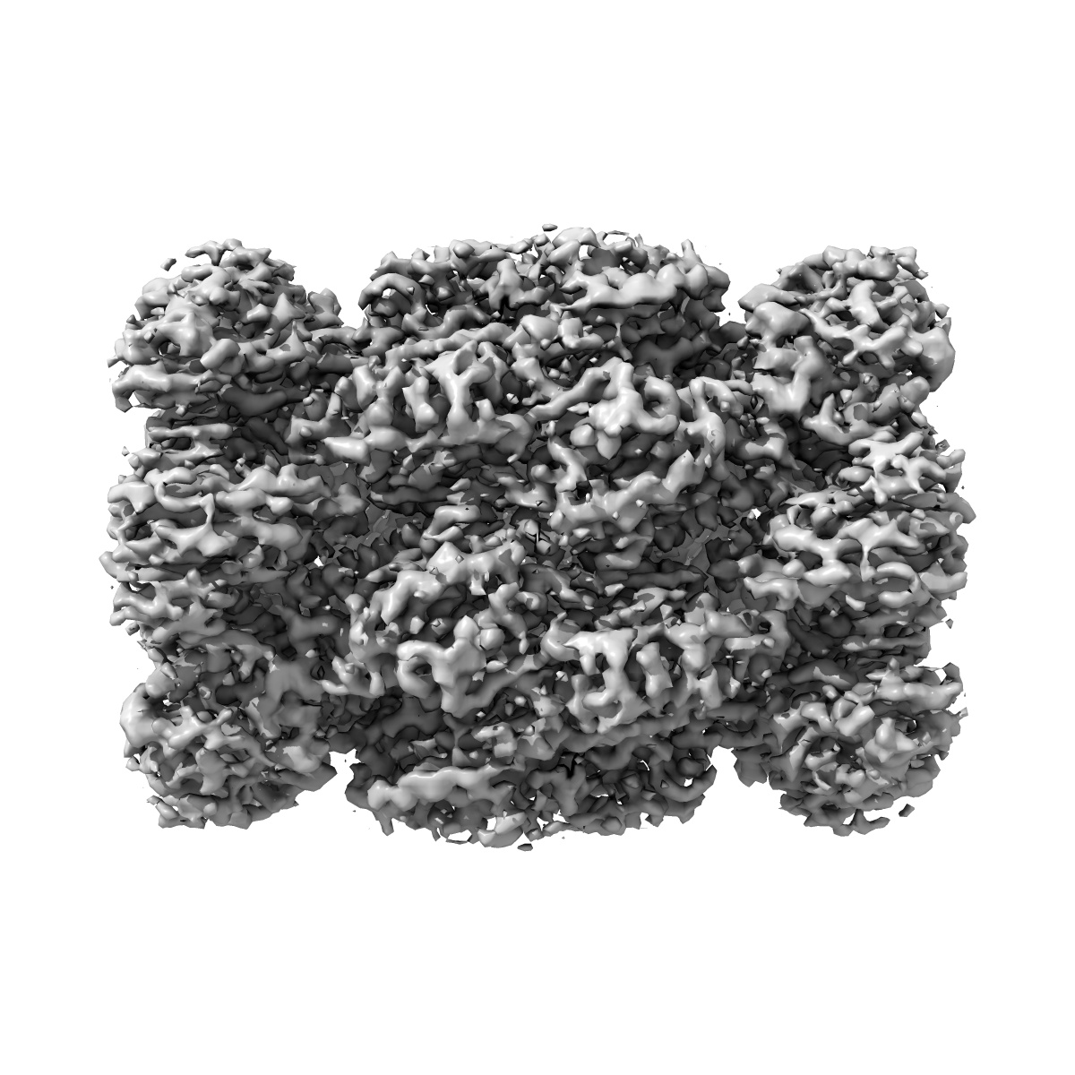
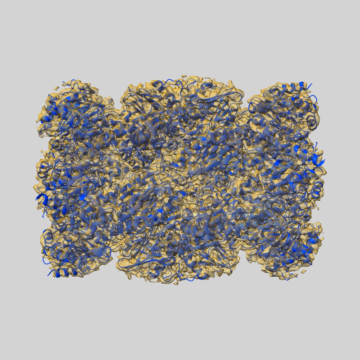
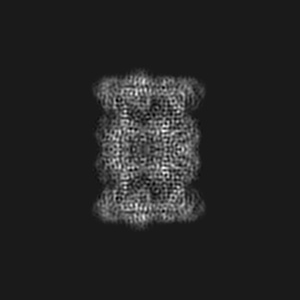
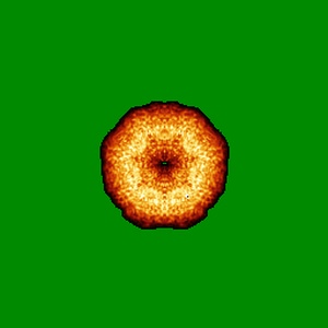
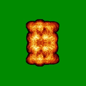
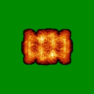
EMD-7010
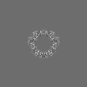
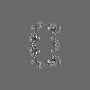
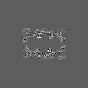
Cryo-EM structure of human immunoproteasome with a novel noncompetitive inhibitor that selectively inhibits activated lymphocytes
EMD-7010
Single-particle3.8 Å
 Deposition: 04/09/2017
Map released: 06/12/2017
Last modified: 13/03/2024
Buffer
pH: 8.0
Vitrification
Cryogen name: ETHANE
Microscope: JEOL 3200FS
Illumination mode: FLOOD BEAM
Imaging mode: BRIGHT FIELD
Electron source: FIELD EMISSION GUN
Acceleration voltage: 300 kV
Illumination mode: FLOOD BEAM
Imaging mode: BRIGHT FIELD
Electron source: FIELD EMISSION GUN
Acceleration voltage: 300 kV
Image Recording
[1]
Final
reconstruction
Resolution: 3.8
Å
(
BY AUTHOR)
Resolution method: FSC 0.143 CUT-OFF
Number of images used: 75017
Resolution method: FSC 0.143 CUT-OFF
Number of images used: 75017
Software
[1]
| Name | Version | Details |
|---|---|---|
| RELION | 1.4 | - |
Startup model
[1]
Type:
NONE
⦨ Initial angle
assignment
Type:
ANGULAR RECONSTITUTION
⦩ Final angle assignment
Type:
ANGULAR RECONSTITUTION
Format: CCP4
Data type: IMAGE STORED AS FLOATING POINT NUMBER (4 BYTES)
Annotation details: Human immunoproteasome with a novel noncompetitive inhibitor
Data type: IMAGE STORED AS FLOATING POINT NUMBER (4 BYTES)
Annotation details: Human immunoproteasome with a novel noncompetitive inhibitor
⬡ Geometry
| X | Y | Z | |
|---|---|---|---|
| Dimensions | 256 | 256 | 256 |
| Origin | 0 | 0 | 0 |
| Spacing | 256 | 256 | 256 |
| Voxel size | 1.2 Å | 1.2 Å | 1.2 Å |
Contour list
| Primary | Level | Source |
|---|---|---|
| True | 0.0383 | AUTHOR |