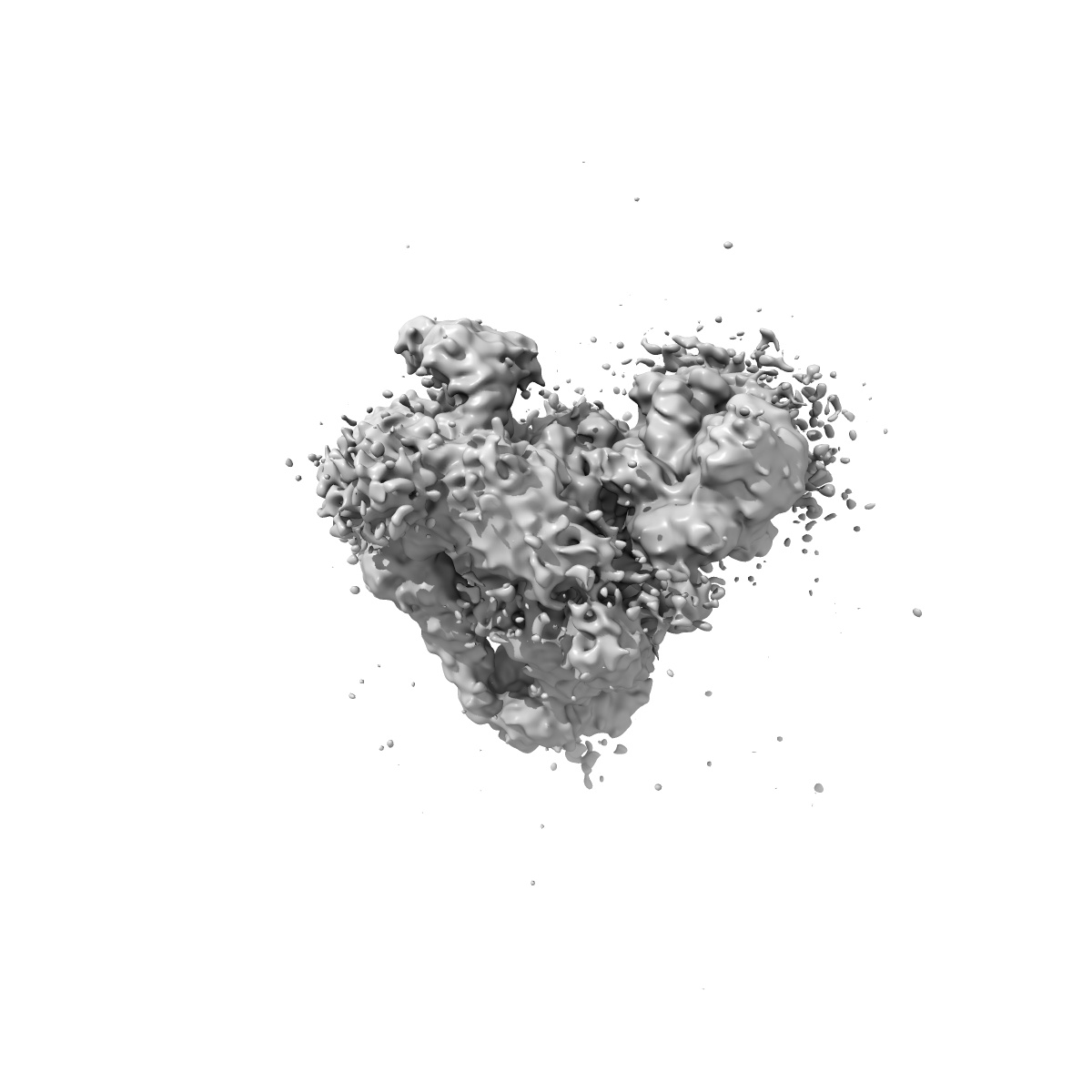
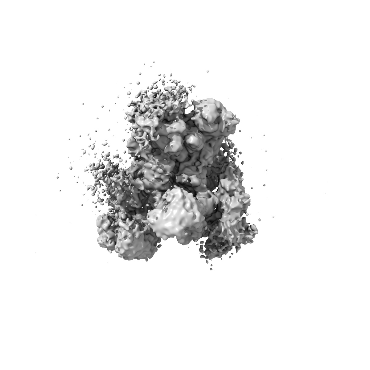
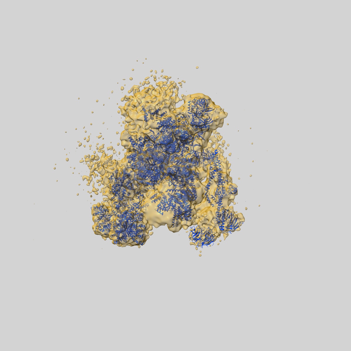
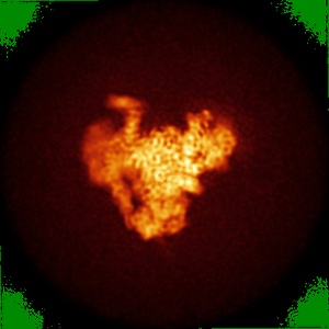
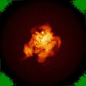
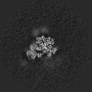
EMD-4057
Structure of the yeast spliceosome immediately after branching. 3D class containing helicase module.
EMD-4057
Single-particle10.0 Å
 Deposition: 12/08/2016
Map released: 31/08/2016
Last modified: 11/12/2019
Concentration: 0.3
mg/mL
Buffer
pH: 7.8
Buffer components [3]:
Buffer components [3]:
| Name | Formula | Concentration | ChEBI |
|---|---|---|---|
| Hepes.KOH pH 7.8 | - | 20.0 millimolar | - |
| Potassium chloride | KCl | 75.0 millimolar | |
| EDTA | - | 250.0 micromolar | - |
Grid
Mesh: 400
Model: Quantifoil R2/2
Material: COPPER
Model: Quantifoil R2/2
Material: COPPER
Pretreatment
Support Film [1]
| Material | Topology | Thickness |
|---|---|---|
| CARBON | CONTINUOUS | 6.0 nm |
Vitrification
Cryogen name: ETHANE
Chamber humidity: 100%
Chamber temperature: 277 K
Instrument: FEI VITROBOT MARK III
Details: 3 microlitres sample were applied to the grid, left for 30 seconds and then blotted for 2.5-3.0 seconds before plunging..
Chamber humidity: 100%
Chamber temperature: 277 K
Instrument: FEI VITROBOT MARK III
Details: 3 microlitres sample were applied to the grid, left for 30 seconds and then blotted for 2.5-3.0 seconds before plunging..
Microscope: FEI TITAN KRIOS
Illumination mode: FLOOD BEAM
Imaging mode: BRIGHT FIELD
Electron source: FIELD EMISSION GUN
Acceleration voltage: 300 kV
Nominal defocus: 0.5 µm - 4.0 µm
Nominal magnification: 81000.0
Calibrated magnification: 35714.0
Specimen holder model: FEI TITAN KRIOS AUTOGRID HOLDER
Cooling holder cryogen: NITROGEN
Specialist optics
Energy filter
Name:
GIF Quantum
Image Recording
[1]
Detector model:
GATAN K2 QUANTUM (4k x 4k)
Detector mode: SUPER-RESOLUTION
Frames per image: 1-20
Number of real images: 2213
Average exposure time: 0.8 s
Average electron dose per image: 2.0 e/Å2
Details: Total dose: 40 electrons/Angstrom^2 over 16 seconds. 20 movie frames collected at 1.25 frames per second.
Detector mode: SUPER-RESOLUTION
Frames per image: 1-20
Number of real images: 2213
Average exposure time: 0.8 s
Average electron dose per image: 2.0 e/Å2
Details: Total dose: 40 electrons/Angstrom^2 over 16 seconds. 20 movie frames collected at 1.25 frames per second.
Final
reconstruction
Resolution: 10.0
Å
(
BY AUTHOR)
Resolution method: FSC 0.143 CUT-OFF
Number of classed used: 1
Number of images used: 15872
Resolution method: FSC 0.143 CUT-OFF
Number of classed used: 1
Number of images used: 15872
Software
[1]
| Name | Version | Details |
|---|---|---|
| RELION | 1.4 | - |
Startup model
[1]
⦨ Initial angle
assignment
⦩ Final angle assignment
Particle selection
[1]
| Selected | Ref. model | Method | Software | Details |
|---|---|---|---|---|
| 15872 | - | - | - | - |
Final 3D classification
Software
[1]
| Name | Version | Details |
|---|---|---|
| RELION | 1.4 | - |
CTF correction
Software
[1]
| Name | Version | Details |
|---|---|---|
| CTFFIND | 4 | - |
Format: CCP4
Data type: IMAGE STORED AS FLOATING POINT NUMBER (4 BYTES)
Data type: IMAGE STORED AS FLOATING POINT NUMBER (4 BYTES)
⬡ Geometry
| X | Y | Z | |
|---|---|---|---|
| Dimensions | 412 | 412 | 412 |
| Origin | 0 | 0 | 0 |
| Spacing | 412 | 412 | 412 |
| Voxel size | 1.43 Å | 1.43 Å | 1.43 Å |
Contour list
| Primary | Level | Source |
|---|---|---|
| True | 0.006 | EMDB |