Across the world’s forests, deserts, and grasslands, snakes have adapted into highly specialized predators. That kind of precision demands more than just fangs. Each snake species carries venom shaped by millions of years of evolution to shut down vital systems of its prey quickly and efficiently - making a single bite devastatingly effective. Some snake venoms hijack the nervous system, causing instant paralysis. Others rupture blood vessels, trigger internal bleeding, or sabotage clotting mechanisms, throwing the circulatory system into chaos. There are also those that weaken muscles, disrupt metabolism, or even manipulate pain perception, buying the snake time to escape or strike again.
How can a single bite deliver such a range of devastating effects?
Snake venom isn’t a single substance, but a heterogeneous mixture of proteins and peptides, often containing up to a hundred different compounds, each evolved to target a specific part of the victim’s physiology. These components can work simultaneously or sequentially to disable, immobilize, and begin digesting prey. No two snake species have the same venom composition, and even individuals within a species can have variations based on age, diet, or geography. Not every compound in snake venom is meant to kill. While some compounds are directly toxic, others support the venom’s effectiveness by helping it spread, persist, or target specific tissues. The toxic compounds fall into four main categories: neurotoxic, hemotoxic, cytotoxic, and myotoxic, depending on the site of action.
Small proteins. Big impact.
In the world of snake venom, size isn’t everything - and three-finger toxins prove it. Three-finger toxins (3FTx) are a family of non-enzymatic proteins found primarily in the venom of elapid snakes like cobras and mambas. These proteins, typically just 60-74 amino acids long, are named for their distinctive structure: three beta-strand loops, or ‘’fingers’’, that extend from a central core and are stabilised by disulfide bonds. Despite this simple architecture, they have a big impact. They can bind to a wide range of biological targets, from ion channels to enzymes to cell surface receptors. Some act as neurotoxins, disrupting nerve signals by locking onto nicotinic acetylcholine receptors, while others function as cardiotoxins, damaging heart muscle cells by disrupting membrane integrity, which can lead to arrhythmia or cardiac arrest.

Figure 1. Three-dimensional structures of three different classes of three-finger toxins showing disulfide bridges.
A) Long-chain toxin (α-Bungarotoxin (1KFH)); B) Short-chain toxin (Erabutoxin a (1QKE)); C) Non-conventional toxin (Candoxin (1JGK)).
α-Bungarotoxin is one of the best-known members of the three-finger toxin family. This neurotoxin, produced by kraits, targets nicotinic acetylcholine receptors at neuromuscular junctions, the vital connection between nerve and muscle. It binds irreversibly to the acetylcholine binding site, mimicking the neurotransmitter while preventing its action. By blocking this signal, α-Bungarotoxin effectively halts communication between nerve and muscle, leading to rapid paralysis, leaving prey immobilized before it even realizes it has been struck.
Structurally, α-bungarotoxin is a single polypeptide chain of 74 amino acids, stabilized by five disulfide bonds that hold it in a rigid three-finger fold. This allows the toxin to maintain an optimal shape for binding. Unlike acetylcholine, which binds briefly and with limited contact, α-bungarotoxin engages multiple contact points across the receptor’s surface, including the interface between subunits. One of its loop regions extends into the ligand-binding site, mimicking the natural neurotransmitter but forming a far more stable interaction. These multiple contact points, along with a very slow dissociation rate, give the toxin its exceptionally high affinity and near-irreversible binding.
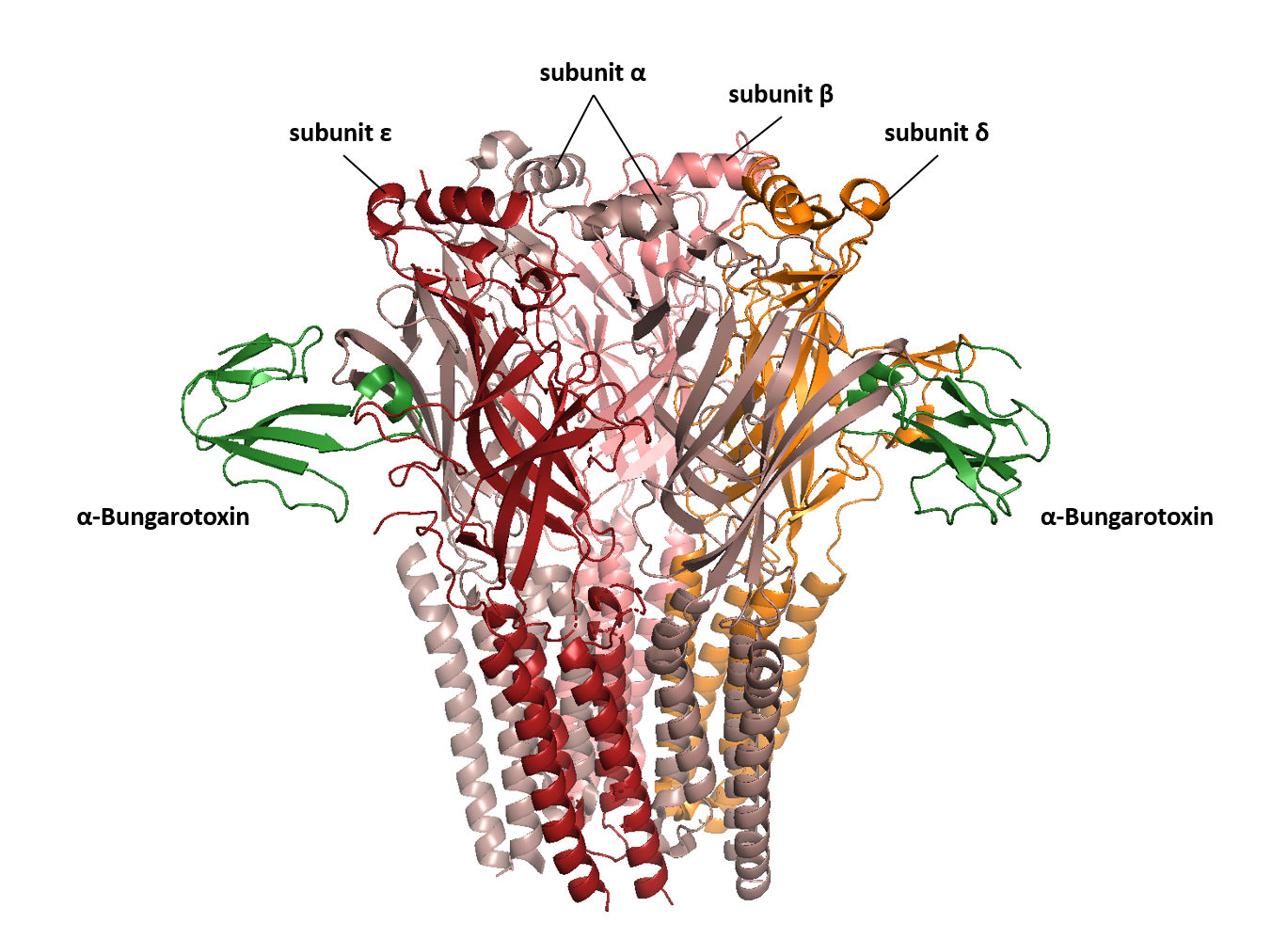
Figure 2. α-Bungarotoxin bound to the α-δ and α-ε interfaces of the human nicotinic acetylcholine receptor (9GU1).
From the elegance of the three-finger fold to the brutal efficiency of paralysis in a single strike, snake venom is designed to kill. And yet its story continues beyond death. Here’s the twist: scientists are turning these deadly compounds into life-saving treatments - painkillers, blood pressure drugs, even cancer therapies – all inspired by snake venom precision. Studying snake venom isn’t just about how snakes kill, it’s about how evolution builds molecular tools and how those same tools might one day be turned toward saving lives rather than ending them.
Did you know?
- Snake bites cause approximately 100,000 deaths worldwide every year.
- About 15-20% of all animal diversity on Earth is venomous.
- The first snake venom toxin to be identified and characterized was crotoxin, a neurotoxin found in the venom of Crotalus durissus terrificus, a South American rattlesnake.
The discovery was made in 1938. - Albert Calmette, a French scientist and physician, is credited with developing the first snake antivenom.
- The only efficient treatment for a snakebite is the administration of a specific antivenom. However, due to the variability in venom composition across snake species and regions, antivenom production and availability remain limited. Most antivenoms are only effective against bites from snakes found in the same geographical region as the venom used to produce them.
Romana Gáborová
About the artwork
Angel Butcher, a Year 12 student at Saffron Walden County High School, found inspiration in the structure of α-bungarotoxin, a snake venom toxin. For the artwork, Angel Butcher created a snake-focused illustration on A3 paper using pencil, fine liner, and markers, incorporating shapes, tones, and detailed patterns to add depth and dimension.
View the artwork in the virtual 2024 PDB Art exhibition.
Structures mentioned in this article
α-Bungarotoxin PDB ID 1KFH
Erabutoxin a PDB ID 1QKE
Candoxin PDB ID 1JGK
Human nicotinic acetylcholine receptor with α-Bungarotoxin PDB ID 9GU1
Sources:
1. The chemistry of snake venom and its medicinal potential
3. Structure, function and evolution of three-finger toxins: Mini proteins with multiple targets
4. Structure and function of α-Bungarotoxin
6. Structures of the human adult muscle-type nicotinic receptor in resting and desensitized states