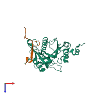
6lph

X-ray diffraction
1.91Å resolution

the Sufu-Fu complex crystal structure

Released:
Source organism: Drosophila melanogaster
Entry authors: Hua L, Geng W

Function and Biology Details

Reaction catalysed:
ATP + a protein = ADP + a phosphoprotein
Biochemical function:
Biological process:
  • not assigned
Cellular component:
  • not assigned

Structure analysis Details

Assembly composition:
hetero dimer (preferred)
PDBe Complex ID:
PDB-CPX-150032 (preferred)
Entry contents:
2 distinct polypeptide molecules
Macromolecules (2 distinct):
Suppressor of fused homolog Chain: A
Molecule details ›
Chain: A
Length: 258 amino acids
Theoretical weight: 29.31 KDa
Source organism: Drosophila melanogaster
Expression system: Escherichia coli
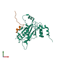
UniProt:
  • Canonical: Q9VG38 (Residues: 1-258; Coverage: 55%)
Gene names: CG6054, Dmel\CG6054, Dmel_CG6054, SU(FU), SUFU, Su(Fu), Su(fu), SuFu, Sufu, d Sufu, dSufu, su(fu), sufu
Sequence domains: Suppressor of fused protein (SUFU)
Serine/threonine-protein kinase fused Chain: B
Molecule details ›
Chain: B
Length: 28 amino acids
Theoretical weight: 3.02 KDa
Source organism: Drosophila melanogaster
Expression system: Escherichia coli
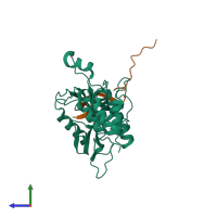
UniProt:
  • Canonical: P23647 (Residues: 363-387; Coverage: 3%)
Gene names: CG6551, fu

Ligands and Environments

No bound ligands
No modified residues

Experiments and Validation Details

Entry percentile scores
X-ray source: SSRF BEAMLINE BL17U
Spacegroup: P21
Unit cell:
a: 36.145Å b: 68.423Å c: 56.144Å
α: 90° β: 97.49° γ: 90°
R-values:
R R work R free
0.151 0.148 0.22
Expression system: Escherichia coli