Xanthine phosphoribosyltransferase
Xanthine-guanine phosphoribosyltransferase catalyses the conversion of guanine, xanthine and, to a lesser extent, hypoxanthine to GMP, XMP and IMP, respectively. E. coli has three PRTases, which are components of the purine salvage pathway. The EC number reaction is shown in the opposite direction to that thought to happen in vitro.
Reference Protein and Structure
- Sequence
-
P0A9M5
 (2.4.2.-, 2.4.2.22)
 (Sequence Homologues)
(PDB Homologues)
(Sequence Homologues)
(PDB Homologues)
- Biological species
-
Escherichia coli K-12 (Bacteria)
- PDB
-
1a95
- XPRTASE FROM E. COLI COMPLEXED WITH MG:CPRPP AND GUANINE
(2.0 Å)


- Catalytic CATH Domains
-
3.40.50.2020
 (see all for 1a95)
- Cofactors
- Magnesium(2+) (1)
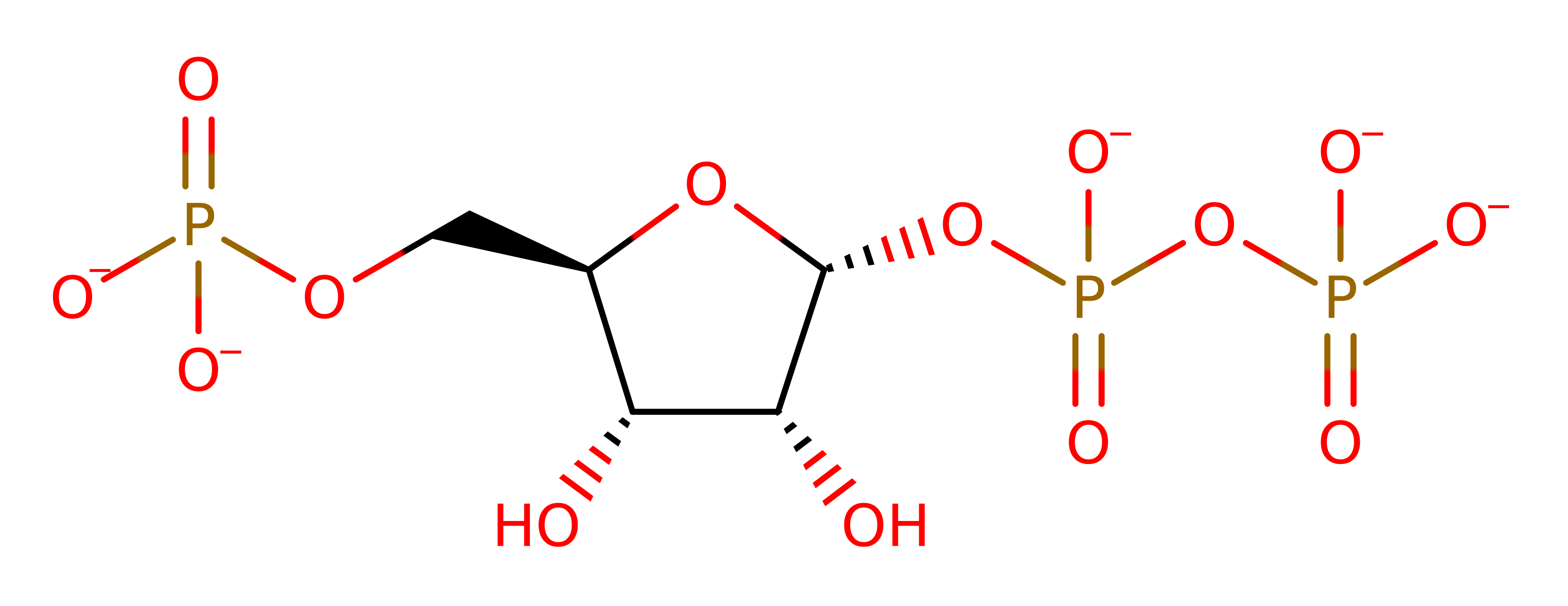
Enzyme Reaction (EC:2.4.2.22)
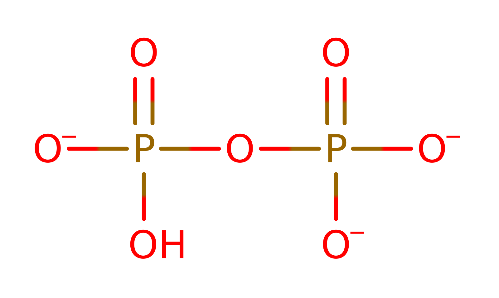
+

→

+
Alternative enzyme names: 5-phospho-alpha-D-ribose-1-diphosphate:xanthine phospho-D-ribosyltransferase, XMP pyrophosphorylase, Xan phosphoribosyltransferase, Xanthosine 5'-phosphate pyrophosphorylase, Xanthylate pyrophosphorylase, Xanthylic pyrophosphorylase, Xanthine-guanine phosphoribosyltransferase,
Enzyme Mechanism
Introduction
alpha-D-5-phosphoribosyl 1-pyrophosphate binds before the purine in an ordered sequential mechanism. Once both substrates are in the active site, Asp92 deprotonates the purine which initiates a nucleophilic attack upon the C2 of the ribose ring of alpha-D-5-phosphoribosyl 1-pyrophosphate in a substitution reaction, eliminating diphosphate.
Catalytic Residues Roles
| UniProt | PDB* (1a95) | ||
| Asp88, Asp89 | Asp88A, Asp89A | Stabilises positive charge on oxocarbonium ion. | electrostatic stabiliser |
| Asp89 | Asp89A | Binds the Mg(II) ion. | electrostatic stabiliser |
| Asp92 | Asp92A | Acts as a general acid/base. Abstracts a proton from the guanine at N7, allowing it to initiate a nucleophilic attack on the ribose ring. | proton shuttle (general acid/base) |
*PDB label guide - RESx(y)B(C) - RES: Residue Name; x: Residue ID in PDB file;
y: Residue ID in PDB sequence if different from PDB file; B: PDB Chain;
C: Biological Assembly Chain if different from PDB. If label is "Not Found" it means this residue is not found in the reference PDB.
Chemical Components
References
- Roy S et al. (2015), Mol Biochem Parasitol, 204, 111-120. Kinetic mechanism of Plasmodium falciparum hypoxanthine-guanine-xanthine phosphoribosyltransferase. DOI:10.1016/j.molbiopara.2016.02.006. PMID:26902413.
- Ullman B et al. (2010), Int J Biochem Cell Biol, 42, 253-262. Acidic residues in the purine binding site govern the 6-oxopurine specificity of the Leishmania donovani xanthine phosphoribosyltransferase. DOI:10.1016/j.biocel.2009.10.020. PMID:19861168.
- Canyuk B et al. (2001), Biochemistry, 40, 2754-2765. The Role for an Invariant Aspartic Acid in Hypoxanthine Phosphoribosyltransferases Is Examined Using Saturation Mutagenesis, Functional Analysis, and X-ray Crystallography†. DOI:10.1021/bi001195q. PMID:11258886.
- Shi W et al. (1999), Nat Struct Biol, 6, 588-593. The 2.0 A structure of human hypoxanthine-guanine phosphoribosyltransferase in complex with a transition-state analog inhibitor. DOI:10.1038/9376. PMID:10360366.
- Héroux A et al. (1999), Biochemistry, 38, 14495-14506. Crystal Structure ofToxoplasma gondiiHypoxanthine-Guanine Phosphoribosyltransferase with XMP, Pyrophosphate, and Two Mg2+Ions Bound: Insights into the Catalytic Mechanism†,‡. DOI:10.1021/bi990508i. PMID:10545171.
- Vos S et al. (1998), J Mol Biol, 282, 875-889. Structures of free and complexed forms of Escherichia coli xanthine-guanine phosphoribosyltransferase1 1Edited by R. Huber. DOI:10.1006/jmbi.1998.2051. PMID:9743633.
- Vos S et al. (1997), Biochemistry, 36, 4125-4134. Crystal Structure ofEscherichia coliXanthine Phosphoribosyltransferase†. DOI:10.1021/bi962640d. PMID:9100006.

Catalytic Residues Roles
| Residue | Roles |
|---|---|
| Asp92A | proton shuttle (general acid/base) |
| Asp88A | electrostatic stabiliser |
| Asp89A | electrostatic stabiliser |