 |
PDBsum entry 6tth

|
|
|
|
|
|

|

|

|

|
|

|
|

|
|

|
|

|
|

|
|

|
|
|
 |

|

|
|
|
|
|
|
|
|
|
|
|
|
|
|
|
|
|
|
|
|
|
|
|
|
|
|
|
|
|
|
|
|
|
|
|
Cytosolic protein
|
PDB id
|
|
|
|
6tth
|
|
|
|
|
|
|
|
|
|
|
|
|
|
|

|

|

|
|
|
|
|
|
|
PDB id:
|
|
|
|
| Name: |
|
Cytosolic protein
|
|
|
Title:
|
|
Pkm2 in complex with l-threonine
|
|
Structure:
|
|
Pyruvate kinase pkm. Chain: a, b, c, d. Synonym: cytosolic thyroid hormone-binding protein,cthbp,opa- interacting protein 3,oip-3,pyruvate kinase 2/3,pyruvate kinase muscle isozyme,thyroid hormone-binding protein 1,thbp1,tumor m2-pk, p58. Engineered: yes
|
|
Source:
|
|
Homo sapiens. Human. Organism_taxid: 9606. Gene: pkm, oip3, pk2, pk3, pkm2. Expressed in: escherichia coli. Expression_system_taxid: 562
|
|
Authors:
|
|
M.Saur,M.J.Hartshorn,J.Dong,J.Reeks,G.Bunkoczi,H.Jhoti,P.A.Williams
|
|
Key ref:
|
|
M.Saur
et al.
(2020).
Fragment-based drug discovery using cryo-EM.
Drug Discov Today,
25,
485-490.
PubMed id:
DOI:
|
|
|
Date:
|
|
|
27-Dec-19
|
Release date:
|
15-Jan-20
|
|
|
|
|
|
|
PROCHECK
|

|
|
|
|
|
Headers
|
|
|
|
References
|
|
|
|
|
|

|
|
P14618
(KPYM_HUMAN) -
Pyruvate kinase PKM from Homo sapiens
|
|
|
|
Seq:
Struc:
|
|
|
|
531 a.a.
410 a.a.
|
|

|

|
|
|
|
|
|
|
|
|
|
|
|
Key: |
 |
PfamA domain |
|
|
 |
Secondary structure |
|
|
|
|
|
|
|
|
|
|
|
|
|
Enzyme class 2:
|
|
E.C.2.7.1.40
- pyruvate kinase.
|
|
|
|
|
|
|
Reaction:
|
|
pyruvate + ATP = phosphoenolpyruvate + ADP + H+
|
|
|
|
|
|
 pyruvate
|
+
|
ATP
Bound ligand (Het Group name = )
matches with 55.56% similarity
|
=
|
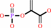 phosphoenolpyruvate
|
+
|
 ADP
ADP
|
+
|
H(+)
Bound ligand (Het Group name = )
matches with 42.86% similarity
|
|
|
|
|
|
|
|
|
|
Enzyme class 3:
|
|
E.C.2.7.10.2
- non-specific protein-tyrosine kinase.
|
|
|
|
|
|
|
Reaction:
|
|
L-tyrosyl-[protein] + ATP = O-phospho-L-tyrosyl-[protein] + ADP + H+
|
|
|
|
|
|
L-tyrosyl-[protein]
|
+
|
 ATP
ATP
|
=
|
O-phospho-L-tyrosyl-[protein]
|
+
|
ADP
|
+
|
H(+)
|
|
|
|
|
|
|
|
|
|
Enzyme class 4:
|
|
E.C.2.7.11.1
- non-specific serine/threonine protein kinase.
|
|
|
|
|
|
|
Reaction:
|
|
|
1.
|
L-seryl-[protein] + ATP = O-phospho-L-seryl-[protein] + ADP + H+
|
|
2.
|
L-threonyl-[protein] + ATP = O-phospho-L-threonyl-[protein] + ADP + H+
|
|
|
|
|
|
|
L-seryl-[protein]
|
+
|
ATP
|
=
|
O-phospho-L-seryl-[protein]
|
+
|
ADP
|
+
|
H(+)
|
|
|
|
|
|
|
L-threonyl-[protein]
|
+
|
ATP
|
=
|
O-phospho-L-threonyl-[protein]
|
+
|
ADP
|
+
|
H(+)
|
|
|
|
|
|
|
|
|
|
|
|
|
Note, where more than one E.C. class is given (as above), each may
correspond to a different protein domain or, in the case of polyprotein
precursors, to a different mature protein.
|
|
|
|
Molecule diagrams generated from .mol files obtained from the
KEGG ftp site
|
|
|
|
|
|
|
|
|
|
|
|
|
|
 |
|
|
|
|
|
|
| |
|
|
| |
|
DOI no:
|
Drug Discov Today
25:485-490
(2020)
|
|
PubMed id:
|
|
|
|
|
|
| |
|
Fragment-based drug discovery using cryo-EM.
|
|
M.Saur,
M.J.Hartshorn,
J.Dong,
J.Reeks,
G.Bunkoczi,
H.Jhoti,
P.A.Williams.
|
|
|
|
|
| |
ABSTRACT
|
|
|
|
| |
|
|
Recent advances in electron cryo-microscopy (cryo-EM) structure determination
have pushed the resolutions obtainable by the method into the range widely
considered to be of utility for drug discovery. Here, we review the use of
cryo-EM in fragment-based drug discovery (FBDD) based on in-house method
development. We demonstrate not only that cryo-EM can reveal details of the
molecular interactions between fragments and a protein, but also that the
current reproducibility, quality, and throughput are compatible with FBDD. We
exemplify this using the test system β-galactosidase (Bgal) and the oncology
target pyruvate kinase 2 (PKM2).
|
|
|
|
|
|
|
|
|
|
|
|
|
|
|
|
|
|
 |
 |
 |
|


 Links
Links


