 |
PDBsum entry 2ct8

|
|
|
|
|
 |
Contents |
 |
|
|
|
|
|
|
|
|
|
|
|
|
|
|
* Residue conservation analysis
|
|
|
|
|
|
PDB id:
|
|
|
|
| Name: |
|
Ligase/RNA
|
|
|
Title:
|
|
Crystal structure of aquifex aeolicus methionyl-tRNA synthetase complexed with tRNA(met) and methionyl-adenylate anologue
|
|
Structure:
|
|
RNA (74-mer). Chain: c, d. Engineered: yes. Methionyl-tRNA synthetase. Chain: a, b. Synonym: methionine--tRNA ligase, metrs. Engineered: yes
|
|
Source:
|
|
Synthetic: yes. Aquifex aeolicus. Organism_taxid: 63363. Expressed in: escherichia coli. Expression_system_taxid: 562.
|
|
Biol. unit:
|
|
 Dimer (from
)
Dimer (from
)
|
|
Resolution:
|
|
|
2.70Å
|
R-factor:
|
0.217
|
R-free:
|
0.272
|
|
|
Authors:
|
|
K.Nakanishi,Y.Ogiso,T.Nakama,S.Fukai,O.Nureki,Riken Structural Genomics/proteomics Initiative (Rsgi)
|
Key ref:

|
|
K.Nakanishi
et al.
(2005).
Structural basis for anticodon recognition by methionyl-tRNA synthetase.
Nat Struct Biol,
12,
931-932.
PubMed id:
DOI:

|
|
|
Date:
|
|
|
23-May-05
|
Release date:
|
20-Sep-05
|
|
|
|
|
|
|
PROCHECK
|

|
|
|
|
|
Headers
|
|
|
|
References
|
|
|
|
|
|

|
|
O67298
(SYM_AQUAE) -
Methionine--tRNA ligase from Aquifex aeolicus (strain VF5)
|
|
|
|
Seq:
Struc:
|
|
|
|
497 a.a.
465 a.a.
|
|

|

|

|
|
|
|
|
|
|
|
|
|
|
Key: |
 |
PfamA domain |
|
|
 |
Secondary structure |
|
 |
CATH domain |
|
|
|
|
|
|
|
|

|
|
|
|
G-G-C-G-G-C-G-U-A-G-C-U-C-A-G-C-U-G-G-U-C-A-G-A-G-C-G-G-G-G-A-U-C-U-C-A-U-A-A-
74 bases
|
|
|
|
G-G-C-G-G-C-G-U-A-G-C-U-C-A-G-C-U-G-G-U-C-A-G-A-G-C-G-G-G-G-A-U-C-U-C-A-U-A-A-
74 bases
|
|
|
|
|
|
|
|
|
|
|
|
|
|
Enzyme class:
|
|
E.C.6.1.1.10
- methionine--tRNA ligase.
|
|
|
|
|
|
|
Reaction:
|
|
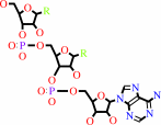
tRNA(Met) + L-methionine + ATP = L-methionyl-tRNA(Met) + AMP + diphosphate
|
|
|
|
|
|
 tRNA(Met)
|
+
|
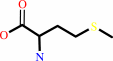 L-methionine
|
+
|
 ATP
ATP
|
=
|
L-methionyl-tRNA(Met)
Bound ligand (Het Group name = )
matches with 54.29% similarity
|
+
|
 AMP
AMP
|
+
|
 diphosphate
|
|
|
|
|
|
|
|
|
|
|
|
|
Molecule diagrams generated from .mol files obtained from the
KEGG ftp site
|
|
|
|
|
|
|
|
|
|
|
|
|
|
 |
|
|
|
|
|
|
| |
|
|
| |
|
DOI no:
|
Nat Struct Biol
12:931-932
(2005)
|
|
PubMed id:
|
|
|
|
|
|
| |
|
Structural basis for anticodon recognition by methionyl-tRNA synthetase.
|
|
K.Nakanishi,
Y.Ogiso,
T.Nakama,
S.Fukai,
O.Nureki.
|
|
|
|
|
| |
ABSTRACT
|
|
|
|
| |
|
|
In the 2.7-A resolution crystal structure of methionyl-tRNA synthetase (MetRS)
in complex with tRNA(Met) and a methionyl-adenylate analog, the tRNA anticodon
loop is distorted to form a triple-base stack comprising C34, A35 and A38. A
tryptophan residue stacks on C34 to extend the triple-base stack. In addition,
C34 forms Watson-Crick-type hydrogen bonds with Arg357. This structure resolves
the longstanding question of how MetRS specifically recognizes tRNA(Met).
|
|
|
|
|
|
| |
Selected figure(s)
|
|
|
|
| |
|
|

|
|

|
|
Figure 1.
Figure 1. The A. aeolicus MetRS−tRNA[m]^Met complex. Green,
the Rossmann fold domain; cyan and dark blue, the CP1 and CP2
insertions, respectively; salmon, the MetRS-specific  -strand
insertion; red, the stem-contact-fold domain; pink, the -strand
insertion; red, the stem-contact-fold domain; pink, the  -helix
bundle domain; yellow, tRNA[m]^Met; stick model, MetSA. -helix
bundle domain; yellow, tRNA[m]^Met; stick model, MetSA.
|
|
Figure 2.
Figure 2. Recognition of the anticodon CAU of tRNA[m]^Met by
MetRS. (a) Conformational change of A. aeolicus tRNA[m]^Met
(yellow) upon binding MetRS, as compared with unbound yeast
tRNA^Phe (blue). (b) Interaction between MetRS and the
tRNA[m]^Met anticodon. Yellow, tRNA[m]^Met nucleotides; light
purple, MetRS residues; dotted lines, hydrogen bonds; red,
oxygen; blue, nitrogen.
|
|
|
|
|
|
| |
The above figures are
reprinted
by permission from Macmillan Publishers Ltd:
Nat Struct Biol
(2005,
12,
931-932)
copyright 2005.
|
|
|
|
|
|
|
|
|
|
|
|
|
|
 |
 |
 |
|
Literature references that cite this PDB file's key reference
|
|
|
| |
PubMed id
|
|
Reference
|
|
|
|
|
|
T.E.Jones,
R.W.Alexander,
and
T.Pan
(2011).
Misacylation of specific nonmethionyl tRNAs by a bacterial methionyl-tRNA synthetase.
|
| |
Proc Natl Acad Sci U S A,
108,
6933-6938.
|
|
|
|
|
|
|
T.Osawa,
S.Kimura,
N.Terasaka,
H.Inanaga,
T.Suzuki,
and
T.Numata
(2011).
Structural basis of tRNA agmatinylation essential for AUA codon decoding.
|
| |
Nat Struct Mol Biol,
18,
1275-1280.
|
|
|
PDB codes:
|
|
|
|
|
|
|
|
H.Ingvarsson,
and
T.Unge
(2010).
Flexibility and communication within the structure of the Mycobacterium smegmatis methionyl-tRNA synthetase.
|
| |
FEBS J,
277,
3947-3962.
|
|
|
PDB codes:
|
|
|
|
|
|
|
|
S.Havrylenko,
R.Legouis,
B.Negrutskii,
and
M.Mirande
(2010).
Methionyl-tRNA synthetase from Caenorhabditis elegans: a specific multidomain organization for convergent functional evolution.
|
| |
Protein Sci,
19,
2475-2484.
|
|
|
|
|
|
|
E.Schmitt,
I.C.Tanrikulu,
T.H.Yoo,
M.Panvert,
D.A.Tirrell,
and
Y.Mechulam
(2009).
Switching from an induced-fit to a lock-and-key mechanism in an aminoacyl-tRNA synthetase with modified specificity.
|
| |
J Mol Biol,
394,
843-851.
|
|
|
PDB codes:
|
|
|
|
|
|
|
|
K.Nakanishi,
L.Bonnefond,
S.Kimura,
T.Suzuki,
R.Ishitani,
and
O.Nureki
(2009).
Structural basis for translational fidelity ensured by transfer RNA lysidine synthetase.
|
| |
Nature,
461,
1144-1148.
|
|
|
PDB codes:
|
|
|
|
|
|
|
|
M.Konno,
T.Sumida,
E.Uchikawa,
Y.Mori,
T.Yanagisawa,
S.Sekine,
and
S.Yokoyama
(2009).
Modeling of tRNA-assisted mechanism of Arg activation based on a structure of Arg-tRNA synthetase, tRNA, and an ATP analog (ANP).
|
| |
FEBS J,
276,
4763-4779.
|
|
|
PDB codes:
|
|
|
|
|
|
|
|
K.Yura
(2008).
[Trial to predict interactions between proteins and biomolecules based on their three-dimensional structures]
|
| |
Yakugaku Zasshi,
128,
1547-1555.
|
|
|
|
|
|
|
P.F.Agris
(2008).
Bringing order to translation: the contributions of transfer RNA anticodon-domain modifications.
|
| |
EMBO Rep,
9,
629-635.
|
|
|
|
|
|
|
A.Ghosh,
and
S.Vishveshwara
(2007).
A study of communication pathways in methionyl- tRNA synthetase by molecular dynamics simulations and structure network analysis.
|
| |
Proc Natl Acad Sci U S A,
104,
15711-15716.
|
|
|
|
|
|
|
I.A.Vasil'eva,
and
N.A.Moor
(2007).
Interaction of aminoacyl-tRNA synthetases with tRNA: general principles and distinguishing characteristics of the high-molecular-weight substrate recognition.
|
| |
Biochemistry (Mosc),
72,
247-263.
|
|
|
|
|
|
|
L.M.Wadley,
K.S.Keating,
C.M.Duarte,
and
A.M.Pyle
(2007).
Evaluating and learning from RNA pseudotorsional space: quantitative validation of a reduced representation for RNA structure.
|
| |
J Mol Biol,
372,
942-957.
|
|
|
|
|
|
|
M.E.Budiman,
M.H.Knaggs,
J.S.Fetrow,
and
R.W.Alexander
(2007).
Using molecular dynamics to map interaction networks in an aminoacyl-tRNA synthetase.
|
| |
Proteins,
68,
670-689.
|
|
|
|
|
|
The most recent references are shown first.
Citation data come partly from CiteXplore and partly
from an automated harvesting procedure. Note that this is likely to be
only a partial list as not all journals are covered by
either method. However, we are continually building up the citation data
so more and more references will be included with time.
Where a reference describes a PDB structure, the PDB
codes are
shown on the right.
|
|


 Links
Links


