Function and Biology

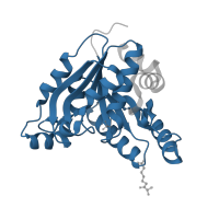
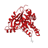
Crystal structure of Mycobacterium tuberculosis dethiobiotin synthetase complexed with 7-Keto 8-aminopelargonic acid

Source organism: Mycobacterium tuberculosis
Biochemical function: metal ion binding
Biological process: biotin biosynthetic process
Cellular component: cytosol

EC 6.3.3.3: Dethiobiotin synthase

Reaction catalysed:
ATP + 7,8-diaminononanoate + CO(2) = ADP + phosphate + dethiobiotin
Systematic name:
7,8-diaminononanoate:carbon-dioxide cyclo-ligase (ADP-forming)
Alternative Name(s):
  • DTB synthetase
  • Desthiobiotin synthase

Sequence family

Pfam Protein family (Pfam)
PF13500
Domain description: AAA domain
Occurring in:
  1. Dethiobiotin synthetase BioD
The deposited structure of PDB entry 3fmi contains 4 copies of Pfam domain PF13500 (AAA domain) in Dethiobiotin synthetase BioD. Showing 1 copy in chain A.

InterPro InterPro annotations
IPR004472
Domain description: Dethiobiotin synthase BioD
Occurring in:
  1. Dethiobiotin synthetase BioD
IPR027417
Domain description: P-loop containing nucleoside triphosphate hydrolase
Occurring in:
  1. Dethiobiotin synthetase BioD
IPR036698
Domain description: TM1070-like superfamily
Occurring in:
  1. Dethiobiotin synthetase BioD

Structure domain

CATH CATH domain
3.40.50.300
Class: Alpha Beta
Architecture: 3-Layer(aba) Sandwich
Topology: Rossmann fold
Homology: P-loop containing nucleotide triphosphate hydrolases
Occurring in:
  1. Dethiobiotin synthetase BioD
The deposited structure of PDB entry 3fmi contains 4 copies of CATH domain 3.40.50.300 (Rossmann fold) in Dethiobiotin synthetase BioD. Showing 1 copy in chain A.