


Function and Biology Details
Biochemical function:
Biological process:
Cellular component:
Structure analysis Details
Assemblies composition:
Assembly name:
DNA mismatch repair protein HSM3 (preferred)
PDBe Complex ID:
PDB-CPX-153616 (preferred)
Entry contents:
1 distinct polypeptide molecule
Macromolecule:
DNA mismatch repair protein HSM3
Molecule details ›
Chains: A, B
Length: 500 amino acids
Theoretical weight: 57.77 KDa
Source organism: Saccharomyces cerevisiae S288C
Expression system: Escherichia coli
UniProt:
Sequence domains:
Length: 500 amino acids
Theoretical weight: 57.77 KDa
Source organism: Saccharomyces cerevisiae S288C
Expression system: Escherichia coli
UniProt:
- Canonical:
 P38348 (Residues: 1-480; Coverage: 100%)
Sequence domains:
- DNA mismatch repair protein HSM3, N terminal domain
- DNA mismatch repair protein HSM3, C terminal domain
Ligands and Environments
No bound ligands
No modified residues
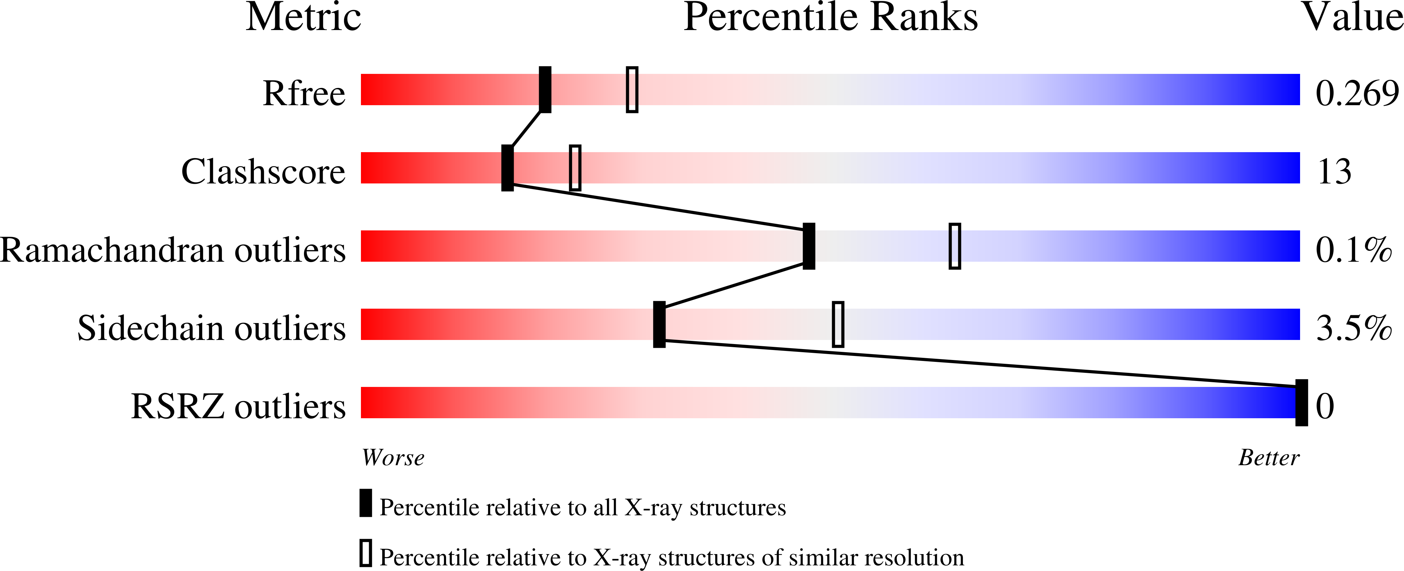
Experiments and Validation Details

X-ray source:
SPRING-8 BEAMLINE BL44XU
Spacegroup:
P21
Expression system: Escherichia coli