 |
PDBsum entry 6b5e
|
|

|
|
PDB id:
|
|
|
|
| Name: |
|
Transferase
|
|
|
Title:
|
|
Mycobacterium tuberculosis rmla in complex with dtdp-glucose
|
|
Structure:
|
|
Glucose-1-phosphate thymidylyltransferase. Chain: a, b, c, d, e, f, g, h. Synonym: dtdp-glucose pyrophosphorylase,dtdp-glucose synthase. Engineered: yes
|
|
Source:
|
|
Mycobacterium tuberculosis (strain atcc 25618 / h37rv). Organism_taxid: 83332. Strain: atcc 25618 / h37rv. Gene: rmla, rfba, rv0334. Expressed in: escherichia coli bl21(de3). Expression_system_taxid: 469008
|
|
Resolution:
|
|
|
1.85Å
|
R-factor:
|
0.174
|
R-free:
|
0.218
|
|
|
Authors:
|
|
H.A.Brown,H.M.Holden
|
|
Key ref:
|
|
H.A.Brown
et al.
(2018).
The structure of glucose-1-phosphate thymidylyltransferase from Mycobacterium tuberculosis reveals the location of an essential magnesium ion in the RmlA-type enzymes.
Protein Sci,
27,
441-450.
PubMed id:
|
|
|
Date:
|
|
|
29-Sep-17
|
Release date:
|
21-Feb-18
|
|
|
|
|
|
|
PROCHECK
|

|
|
|
|
|
Headers
|
|
|
|
References
|
|
|
|
|
|

|
|
P9WH13
(RMLA_MYCTU) -
Glucose-1-phosphate thymidylyltransferase from Mycobacterium tuberculosis (strain ATCC 25618 / H37Rv)
|
|
|
|
Seq:
Struc:
|
|
|
|
288 a.a.
283 a.a.
|
|

|

|

|
|
|
|
|
|
|
|
|
|
|
Key: |
 |
PfamA domain |
|
|
 |
Secondary structure |
|
|
|
|
|
|
|
|
|
|
|
|
|
Enzyme class:
|
|
E.C.2.7.7.24
- glucose-1-phosphate thymidylyltransferase.
|
|
|
|
|
|
|
Pathway:
|
|
6-Deoxyhexose Biosynthesis
|
|
|
|
|
|
Reaction:
|
|
dTTP + alpha-D-glucose 1-phosphate + H+ = dTDP-alpha-D-glucose + diphosphate
|
|
|
|
|
|
 dTTP
dTTP
|
+
|
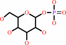 alpha-D-glucose 1-phosphate
|
+
|
H(+)
|
=
|
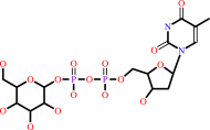 dTDP-alpha-D-glucose
|
+
|
diphosphate
Bound ligand (Het Group name = )
corresponds exactly
|
|
|
|
|
|
|
|
|
|
|
|
|
Molecule diagrams generated from .mol files obtained from the
KEGG ftp site
|
|
|
|
|
|
|
|
|
|
|
|
|
|
 |
|
|
|
|
|
|
| |
|
|
| |
|
|
Protein Sci
27:441-450
(2018)
|
|
PubMed id:
|
|
|
|
|
|
| |
|
The structure of glucose-1-phosphate thymidylyltransferase from Mycobacterium tuberculosis reveals the location of an essential magnesium ion in the RmlA-type enzymes.
|
|
H.A.Brown,
J.B.Thoden,
P.A.Tipton,
H.M.Holden.
|
|
|
|
|
| |
ABSTRACT
|
|
|
|
| |
|
|
Tuberculosis, caused by the bacterium Mycobacterium tuberculosis, continues to
be a major threat to populations worldwide. Whereas the disease is treatable,
the drug regimen is arduous at best with the use of four antimicrobials over a
six-month period. There is clearly a pressing need for the development of new
therapeutics. One potential target for structure-based drug design is the enzyme
RmlA, a glucose-1-phosphate thymidylyltransferase. This enzyme catalyzes the
first step in the biosynthesis of l-rhamnose, which is a deoxysugar critical for
the integrity of the bacterium's cell wall. Here, we report the X-ray structures
of M. tuberculosis RmlA in complex with either dTTP or dTDP-glucose to 1.6 Å
and 1.85 Å resolution, respectively. In the RmlA/dTTP complex, two magnesium
ions were observed binding to the nucleotide, both ligated in octahedral
coordination spheres. In the RmlA/dTDP-glucose complex, only a single magnesium
ion was observed. Importantly, for RmlA-type enzymes with known
three-dimensional structures, not one model shows the position of the magnesium
ion bound to the nucleotide-linked sugar. As such, this investigation represents
the first direct observation of the manner in which a magnesium ion is
coordinated to the RmlA product and thus has important ramifications for
structure-based drug design. In the past, molecular modeling procedures have
been employed to derive a three-dimensional model of the M. tuberculosis RmlA
for drug design. The X-ray structures presented herein provide a superior
molecular scaffold for such endeavors in the treatment of one of the world's
deadliest diseases.
|
|
|
|
|
|
|
|
|
|
|
|
|
|
|
|
|
|
 |
 |
 |
|


 Links
Links


