 |
PDBsum entry 1dr1

|
|
|
|
|
|

|

|

|

|
|

|
|

|
|

|
|

|
|
|
 |

|

|
|
|
|
|
|
|
|
|
|
|
|
|
|
|
|
|
|
|
|
|
|
|
|
|
|
|
|
|
|
|
|
|
Oxidoreductase
|
PDB id
|
|
|
|
1dr1
|
|
|
|
|
|
|
|
|
|
|
|
|
|
|

|

|

|
|
|
|
|
|
|
|
 |
Contents |
 |
|
|
|
|
|
|
|
|
|
|
|
|
|
|
* Residue conservation analysis
|
|
|
|
|
|
|
|
|
|
|
Enzyme class:
|
|
E.C.1.5.1.3
- dihydrofolate reductase.
|
|
|
|
|
|
|
Pathway:
|
|
Folate Coenzymes
|
|
|
|
|
|
Reaction:
|
|
(6S)-5,6,7,8-tetrahydrofolate + NADP+ = 7,8-dihydrofolate + NADPH + H+
|
|
|
|
|
|
(6S)-5,6,7,8-tetrahydrofolate
|
+
|
NADP(+)
Bound ligand (Het Group name = )
corresponds exactly
|
=
|
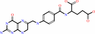 7,8-dihydrofolate
|
+
|
 NADPH
NADPH
|
+
|
H(+)
|
|
|
|
|
|
|
|
|
|
|
|
|
Molecule diagrams generated from .mol files obtained from the
KEGG ftp site
|
|
|
|
|
|
|
|
|
|
|
|
|
|
 |
|
|
|
|
|
|
| |
|
|
| |
|
DOI no:
|
Biochemistry
31:7264-7273
(1992)
|
|
PubMed id:
|
|
|
|
|
|
| |
|
Crystal structure of chicken liver dihydrofolate reductase complexed with NADP+ and biopterin.
|
|
M.A.McTigue,
J.F.Davies,
B.T.Kaufman,
J.Kraut.
|
|
|
|
|
| |
ABSTRACT
|
|
|
|
| |
|
|
The 2.2-A crystal structure of chicken liver dihydrofolate reductase (EC
1.5.1.3, DHFR) has been solved as a ternary complex with NADP+ and biopterin (a
poor substrate). The space group and unit cell are isomorphous with the
previously reported structure of chicken liver DHFR complexed with NADPH and
phenyltriazine [Volz, K. W., Matthews, D. A., Alden, R. A., Freer, S. T.,
Hansch, C., Kaufman, B. T., & Kraut, J. (1982) J. Biol. Chem. 257,
2528-2536]. The structure contains an ordered water molecule hydrogen-bonded to
both hydroxyls of the biopterin dihydroxypropyl group as well as to O4 and N5 of
the biopterin pteridine ring. This water molecule, not observed in previously
determined DHFR structures, is positioned to complete a proposed route for
proton transfer from the side-chain carboxylate of E30 to N5 of the pteridine
ring. Protonation of N5 is believed to occur during the reduction of
dihydropteridine substrates. The positions of the NADP+ nicotinamide and
biopterin pteridine rings are quite similar to the nicotinamide and pteridine
ring positions in the Escherichia coli DHFR.NADP+.folate complex [Bystroff, C.,
Oatley, S. J., & Kraut, J. (1990) Biochemistry 29, 3263-3277], suggesting
that the reduction of biopterin and the reduction of folate occur via similar
mechanisms, that the binding geometry of the nicotinamide and pteridine rings is
conserved between DHFR species, and that the p-aminobenzoylglutamate moiety of
folate is not required for correct positioning of the pteridine ring in
ground-state ternary complexes. Instead, binding of the p-aminobenzoylglutamate
moiety of folate may induce the side chain of residue 31 (tyrosine or
phenylalanine) in vertebrate DHFRs to adopt a conformation in which the opening
to the pteridine binding site is too narrow to allow the substrate to diffuse
away rapidly. A reverse conformational change of residue 31 is proposed to be
required for tetrahydrofolate release.
|
|
|
|
|
|
|
|
|
|
|
|
|
|
|
|
|
|
 |
 |
 |
|
Literature references that cite this PDB file's key reference
|
|
|
| |
PubMed id
|
|
Reference
|
|
|
|
|
|
J.P.Volpato,
B.J.Yachnin,
J.Blanchet,
V.Guerrero,
L.Poulin,
E.Fossati,
A.M.Berghuis,
and
J.N.Pelletier
(2009).
Multiple conformers in active site of human dihydrofolate reductase F31R/Q35E double mutant suggest structural basis for methotrexate resistance.
|
| |
J Biol Chem,
284,
20079-20089.
|
|
|
PDB code:
|
|
|
|
|
|
|
|
Z.Gáspári,
G.Pál,
and
A.Perczel
(2008).
A redesigned genetic code for selective labeling in protein NMR.
|
| |
Bioessays,
30,
772-780.
|
|
|
|
|
|
|
I.V.Khavrutskii,
D.J.Price,
J.Lee,
and
C.L.Brooks
(2007).
Conformational change of the methionine 20 loop of Escherichia coli dihydrofolate reductase modulates pKa of the bound dihydrofolate.
|
| |
Protein Sci,
16,
1087-1100.
|
|
|
|
|
|
|
J.Yuvaniyama,
P.Chitnumsub,
S.Kamchonwongpaisan,
J.Vanichtanankul,
W.Sirawaraporn,
P.Taylor,
M.D.Walkinshaw,
and
Y.Yuthavong
(2003).
Insights into antifolate resistance from malarial DHFR-TS structures.
|
| |
Nat Struct Biol,
10,
357-365.
|
|
|
PDB codes:
|
|
|
|
|
|
|
|
M.Garcia-Viloca,
D.G.Truhlar,
and
J.Gao
(2003).
Reaction-path energetics and kinetics of the hydride transfer reaction catalyzed by dihydrofolate reductase.
|
| |
Biochemistry,
42,
13558-13575.
|
|
|
|
|
|
|
P.Shrimpton,
A.Mullaney,
and
R.K.Allemann
(2003).
Functional role for Tyr 31 in the catalytic cycle of chicken dihydrofolate reductase.
|
| |
Proteins,
51,
216-223.
|
|
|
|
|
|
|
P.Shrimpton,
and
R.K.Allemann
(2002).
Role of water in the catalytic cycle of E. coli dihydrofolate reductase.
|
| |
Protein Sci,
11,
1442-1451.
|
|
|
|
|
|
|
O.A.Santos-Filho,
R.B.de Alencastro,
and
J.D.Figueroa-Villar
(2001).
Homology modeling of wild type and pyrimethamine/cycloguanil-cross resistant mutant type Plasmodium falciparum dihydrofolate reductase. A model for antimalarial chemotherapy resistance.
|
| |
Biophys Chem,
91,
305-317.
|
|
|
|
|
|
|
B.Almås,
K.Toska,
K.Teigen,
V.Groehn,
W.Pfleiderer,
A.Martínez,
T.Flatmark,
and
J.Haavik
(2000).
A kinetic and conformational study on the interaction of tetrahydropteridines with tyrosine hydroxylase.
|
| |
Biochemistry,
39,
13676-13686.
|
|
|
|
|
|
|
G.Rastelli,
W.Sirawaraporn,
P.Sompornpisut,
T.Vilaivan,
S.Kamchonwongpaisan,
R.Quarrell,
G.Lowe,
Y.Thebtaranonth,
and
Y.Yuthavong
(2000).
Interaction of pyrimethamine, cycloguanil, WR99210 and their analogues with Plasmodium falciparum dihydrofolate reductase: structural basis of antifolate resistance.
|
| |
Bioorg Med Chem,
8,
1117-1128.
|
|
|
|
|
|
|
J.D.Szustakowski,
and
Z.Weng
(2000).
Protein structure alignment using a genetic algorithm.
|
| |
Proteins,
38,
428-440.
|
|
|
|
|
|
|
T.Doukov,
J.Seravalli,
J.J.Stezowski,
and
S.W.Ragsdale
(2000).
Crystal structure of a methyltetrahydrofolate- and corrinoid-dependent methyltransferase.
|
| |
Structure,
8,
817-830.
|
|
|
PDB code:
|
|
|
|
|
|
|
|
K.E.Goodwill,
C.Sabatier,
and
R.C.Stevens
(1998).
Crystal structure of tyrosine hydroxylase with bound cofactor analogue and iron at 2.3 A resolution: self-hydroxylation of Phe300 and the pterin-binding site.
|
| |
Biochemistry,
37,
13437-13445.
|
|
|
PDB code:
|
|
|
|
|
|
|
|
A.D.Mesecar,
B.L.Stoddard,
and
D.E.Koshland
(1997).
Orbital steering in the catalytic power of enzymes: small structural changes with large catalytic consequences.
|
| |
Science,
277,
202-206.
|
|
|
PDB codes:
|
|
|
|
|
|
|
|
J.M.Johnson,
E.M.Meiering,
J.E.Wright,
J.Pardo,
A.Rosowsky,
and
G.Wagner
(1997).
NMR solution structure of the antitumor compound PT523 and NADPH in the ternary complex with human dihydrofolate reductase.
|
| |
Biochemistry,
36,
4399-4411.
|
|
|
|
|
|
|
M.R.Sawaya,
and
J.Kraut
(1997).
Loop and subdomain movements in the mechanism of Escherichia coli dihydrofolate reductase: crystallographic evidence.
|
| |
Biochemistry,
36,
586-603.
|
|
|
PDB codes:
|
|
|
|
|
|
|
|
H.Lee,
V.M.Reyes,
and
J.Kraut
(1996).
Crystal structures of Escherichia coli dihydrofolate reductase complexed with 5-formyltetrahydrofolate (folinic acid) in two space groups: evidence for enolization of pteridine O4.
|
| |
Biochemistry,
35,
7012-7020.
|
|
|
PDB codes:
|
|
|
|
|
|
|
|
J.D.Cronk,
J.A.Endrizzi,
and
T.Alber
(1996).
High-resolution structures of the bifunctional enzyme and transcriptional coactivator DCoH and its complex with a product analogue.
|
| |
Protein Sci,
5,
1963-1972.
|
|
|
PDB codes:
|
|
|
|
|
|
|
|
O.Rimet,
M.Chauvet,
M.Dell'Amico,
G.Noat,
and
M.Bourdeaux
(1995).
Variations in fluorescence and enzymic properties of bovine dihydrofolate reductase.NADPH complex during the slow conformational change induced by coenzyme binding.
|
| |
Eur J Biochem,
228,
55-59.
|
|
|
|
|
|
|
W.S.Lewis,
V.Cody,
N.Galitsky,
J.R.Luft,
W.Pangborn,
S.K.Chunduru,
H.T.Spencer,
J.R.Appleman,
and
R.L.Blakley
(1995).
Methotrexate-resistant variants of human dihydrofolate reductase with substitutions of leucine 22. Kinetics, crystallography, and potential as selectable markers.
|
| |
J Biol Chem,
270,
5057-5064.
|
|
|
PDB codes:
|
|
|
|
|
|
|
|
P.L.Cummins,
and
J.E.Gready
(1993).
Computer-aided drug design: a free energy perturbation study on the binding of methyl-substituted pterins and N5-deazapterins to dihydrofolate reductase.
|
| |
J Comput Aided Mol Des,
7,
535-555.
|
|
|
|
|
|
The most recent references are shown first.
Citation data come partly from CiteXplore and partly
from an automated harvesting procedure. Note that this is likely to be
only a partial list as not all journals are covered by
either method. However, we are continually building up the citation data
so more and more references will be included with time.
Where a reference describes a PDB structure, the PDB
code is
shown on the right.
|
|


 Links
Links


