 |
PDBsum entry 2ith

|
|
|
|
|
|

|

|

|

|
|

|
|

|
|

|
|
|
 |

|

|
|
|
|
|
|
|
|
|
|
|
|
|
|
|
|
|
|
|
|
|
|
|
|
|
|
|
|
|
|
|
|
Oxidoreductase
|
PDB id
|
|
|
|
2ith
|
|
|
|
|
|
|
|
|
|
|
|
|
|
|

|

|

|
|
|
|
|
|
|
|
 |
Contents |
 |
|
|
|
|
|
|
|
|
* Residue conservation analysis
|
|
|
|
|
|
|
|
|
|
|
Enzyme class:
|
|
E.C.1.5.1.3
- dihydrofolate reductase.
|
|
|
|
|
|
|
Pathway:
|
|
Folate Coenzymes
|
|
|
|
|
|
Reaction:
|
|
(6S)-5,6,7,8-tetrahydrofolate + NADP+ = 7,8-dihydrofolate + NADPH + H+
|
|
|
|
|
|
(6S)-5,6,7,8-tetrahydrofolate
|
+
|
 NADP(+)
|
=
|
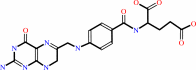 7,8-dihydrofolate
|
+
|
 NADPH
NADPH
|
+
|
H(+)
|
|
|
|
|
|
|
|
|
|
|
|
|
Molecule diagrams generated from .mol files obtained from the
KEGG ftp site
|
|
|
|
|
|
|
|
|
|
|
|
|
|
 |
|
|
|
|
|
|
| |
|
|
| |
|
DOI no:
|
Protein Sci
16:1783-1787
(2007)
|
|
PubMed id:
|
|
|
|
|
|
| |
|
Structure in an extreme environment: NMR at high salt.
|
|
B.Binbuga,
A.F.Boroujerdi,
J.K.Young.
|
|
|
|
|
| |
ABSTRACT
|
|
|
|
| |
|
|
Proteins from halophiles have adapted to challenging environmental conditions
and require salt for their structure and function. How halophilic proteins
adapted to a hypersaline environment is still an intriguing question. It is
important to mimic the physiological conditions of the archae extreme halophiles
when characterizing their enzymes, including structural characterization. The
NMR derived structure of Haloferax volcanii dihydrofolate reductase in 3.5 M
NaCl is presented, and represents the first high salt structure calculated using
NMR data. Structure calculations show that this protein has a solution structure
which is similar to the previously determined crystal structure with a
difference at the N terminus of beta3 and the type of beta-turn connection beta7
and beta8.
|
|
|
|
|
|
| |
Selected figure(s)
|
|
|
|
| |
|
|

|
|
Figure 1.
Figure 1. (A) Backbone superimposition of the 20 lowest energy structures of hvDHFR1, and (B) a representative structure generated
|
|
|
|
|
|
| |
The above figure is
reprinted
by permission from the Protein Society:
Protein Sci
(2007,
16,
1783-1787)
copyright 2007.
|
|
|
|
|
|
|
|
|
|
|
|
|
|
 |
 |
 |
|
Literature references that cite this PDB file's key reference
|
|
|
| |
PubMed id
|
|
Reference
|
|
|
|
|
|
A.F.Boroujerdi,
and
J.K.Young
(2009).
NMR-derived folate-bound structure of dihydrofolate reductase 1 from the halophile Haloferax volcanii.
|
| |
Biopolymers,
91,
140-144.
|
|
|
PDB code:
|
|
|
|
|
|
|
The most recent references are shown first.
Citation data come partly from CiteXplore and partly
from an automated harvesting procedure. Note that this is likely to be
only a partial list as not all journals are covered by
either method. However, we are continually building up the citation data
so more and more references will be included with time.
Where a reference describes a PDB structure, the PDB
code is
shown on the right.
|
|


 Links
Links


