 |
PDBsum entry 1dr3

|
|
|
|
|
|

|

|

|

|
|

|
|

|
|

|
|

|
|

|
|

|
|
|
 |

|

|
|
|
|
|
|
|
|
|
|
|
|
|
|
|
|
|
|
|
|
|
|
|
|
|
|
|
|
|
|
|
|
|
|
|
Oxidoreductase
|
PDB id
|
|
|
|
1dr3
|
|
|
|
|
|
|
|
|
|
|
|
|
|
|

|

|

|
|
|
|
|
|
|
|
 |
Contents |
 |
|
|
|
|
|
|
|
|
|
|
|
|
|
|
* Residue conservation analysis
|
|
|
|
|
|
|
|
|
|
|
Enzyme class:
|
|
E.C.1.5.1.3
- dihydrofolate reductase.
|
|
|
|
|
|
|
Pathway:
|
|
Folate Coenzymes
|
|
|
|
|
|
Reaction:
|
|
(6S)-5,6,7,8-tetrahydrofolate + NADP+ = 7,8-dihydrofolate + NADPH + H+
|
|
|
|
|
|
(6S)-5,6,7,8-tetrahydrofolate
|
+
|
NADP(+)
Bound ligand (Het Group name = )
matches with 95.92% similarity
|
=
|
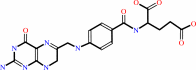 7,8-dihydrofolate
|
+
|
 NADPH
NADPH
|
+
|
H(+)
|
|
|
|
|
|
|
|
|
|
|
|
|
Molecule diagrams generated from .mol files obtained from the
KEGG ftp site
|
|
|
|
|
|
|
|
|
|
|
|
|
|
 |
|
|
|
|
|
|
| |
|
|
| |
|
DOI no:
|
Biochemistry
32:6855-6862
(1993)
|
|
PubMed id:
|
|
|
|
|
|
| |
|
Crystal structures of chicken liver dihydrofolate reductase: binary thioNADP+ and ternary thioNADP+.biopterin complexes.
|
|
M.A.McTigue,
J.F.Davies,
B.T.Kaufman,
J.Kraut.
|
|
|
|
|
| |
ABSTRACT
|
|
|
|
| |
|
|
The role of the 3'-carboxamide substituent of NADPH in the reduction of
pteridine substrates as catalyzed by dihydrofolate reductase (EC 1.5.1.3, DHFR)
has been investigated by determining crystal structures at 2.3 A of chicken
liver DHFR in a binary complex with oxidized thionicotinamide adenine
dinucleotide (thioNADP+) and in a ternary complex with thioNADP+ and biopterin.
These structures are isomorphous with those previously reported for chicken
liver DHFR [Volz, K.W., Matthews, D.A., Alden, R.A., Freer, S. T., Hansch, C.,
Kaufman, B. T., & Kraut, J. (1982) J. Biol. Chem. 257, 2528-2536].
ThioNADPH, which has a 3'-carbothioamide substituent in place of a
3'-carboxamide, functions very poorly as a coenzyme for DHFR [Williams, T. J.,
Lee, T. K., & Dunlap, R. B. (1977) Arch, Biochem. Biophys. 181, 569-579;
Stone, S. R., Mark, A., & Morrison, J. F. (1984) Biochemistry 23,
4340-4346]. Comparisons show that, while NADP+ and NADPH bind to DHFR with the
pyridine ring and 3'-carboxamide coplanar, the thioamide group is twisted by 23
degrees from the pyridine plane in both the binary and ternary complexes. This
twist appears to be due to steric conflict between the thioamide sulfur atom and
both the pyridine ring at C4 and the adjacent protein backbone at Ala-9. It
results in an unfavorably close contact between the sulfur and the biopterin
pteridine ring (0.9 A less than the van der Waals separation) which, on the
basis of the refined structure, greatly destabilizes the binding of
biopterin.(ABSTRACT TRUNCATED AT 250 WORDS)
|
|
|
|
|
|
|
|
|
|
|
|
|
|
|
|
|
|
 |
 |
 |
|
Literature references that cite this PDB file's key reference
|
|
|
| |
PubMed id
|
|
Reference
|
|
|
|
|
|
R.P.Ilagan,
M.Tiso,
D.W.Konas,
C.Hemann,
D.Durra,
R.Hille,
and
D.J.Stuehr
(2008).
Differences in a conformational equilibrium distinguish catalysis by the endothelial and neuronal nitric-oxide synthase flavoproteins.
|
| |
J Biol Chem,
283,
19603-19615.
|
|
|
|
|
|
|
A.Singh,
J.D.Venning,
P.G.Quirk,
G.I.van Boxel,
D.J.Rodrigues,
S.A.White,
and
J.B.Jackson
(2003).
Interactions between transhydrogenase and thio-nicotinamide Analogues of NAD(H) and NADP(H) underline the importance of nucleotide conformational changes in coupling to proton translocation.
|
| |
J Biol Chem,
278,
33208-33216.
|
|
|
PDB codes:
|
|
|
|
|
|
|
|
P.Shrimpton,
A.Mullaney,
and
R.K.Allemann
(2003).
Functional role for Tyr 31 in the catalytic cycle of chicken dihydrofolate reductase.
|
| |
Proteins,
51,
216-223.
|
|
|
|
|
|
|
Y.Q.Shen,
S.Y.Song,
and
Z.J.Lin
(2002).
Structures of D-glyceraldehyde-3-phosphate dehydrogenase complexed with coenzyme analogues.
|
| |
Acta Crystallogr D Biol Crystallogr,
58,
1287-1297.
|
|
|
PDB codes:
|
|
|
|
|
|
|
|
T.Doukov,
J.Seravalli,
J.J.Stezowski,
and
S.W.Ragsdale
(2000).
Crystal structure of a methyltetrahydrofolate- and corrinoid-dependent methyltransferase.
|
| |
Structure,
8,
817-830.
|
|
|
PDB code:
|
|
|
|
|
|
|
|
C.S.Raman,
H.Li,
P.Martásek,
V.Král,
B.S.Masters,
and
T.L.Poulos
(1998).
Crystal structure of constitutive endothelial nitric oxide synthase: a paradigm for pterin function involving a novel metal center.
|
| |
Cell,
95,
939-950.
|
|
|
PDB codes:
|
|
|
|
|
|
|
|
K.K.Koretke,
Z.Luthey-Schulten,
and
P.G.Wolynes
(1996).
Self-consistently optimized statistical mechanical energy functions for sequence structure alignment.
|
| |
Protein Sci,
5,
1043-1059.
|
|
|
|
|
|
The most recent references are shown first.
Citation data come partly from CiteXplore and partly
from an automated harvesting procedure. Note that this is likely to be
only a partial list as not all journals are covered by
either method. However, we are continually building up the citation data
so more and more references will be included with time.
Where a reference describes a PDB structure, the PDB
codes are
shown on the right.
|
|


 Links
Links


