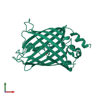
Assemblies
Assembly Name:
Green fluorescent protein
Multimeric state:
monomeric
Accessible surface area:
10546.27 Å2
Buried surface area:
0.0 Å2
Dissociation area:
0
Å2
Dissociation energy (ΔGdiss):
0
kcal/mol
Dissociation entropy (TΔSdiss):
0
kcal/mol
Symmetry number:
1
PDBe Complex ID:
PDB-CPX-154537
Macromolecules
Chain: A
Length: 241 amino acids
Theoretical weight: 27.39 KDa
Source organism: Aequorea victoria
Expression system: Escherichia coli BL21(DE3)
UniProt:
Pfam: Green fluorescent protein
InterPro:
CATH: Green fluorescent protein
Length: 241 amino acids
Theoretical weight: 27.39 KDa
Source organism: Aequorea victoria
Expression system: Escherichia coli BL21(DE3)
UniProt:
- Canonical:
 P42212 (Residues: 2-238; Coverage: 100%)
Pfam: Green fluorescent protein
InterPro:
CATH: Green fluorescent protein


