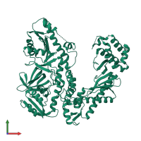
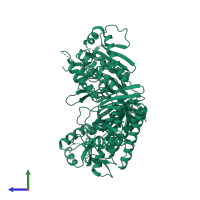
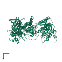
Function and Biology Details
Reaction catalysed:
Deoxynucleoside triphosphate + DNA(n) = diphosphate + DNA(n+1)
Biochemical function:
Biological process:
Cellular component:
- not assigned
Sequence domains:
- DNA-directed DNA polymerase, family B
- DNA polymerase, palm domain superfamily
- DNA polymerase family B, thumb domain
- DNA-directed DNA polymerase, family B, conserved site
- DNA/RNA polymerase superfamily
- DNA-directed DNA polymerase, family B, multifunctional domain
- DNA-directed DNA polymerase, family B, exonuclease domain
- Ribonuclease H superfamily
1 more domain
Structure domains:
Structure analysis Details
Assembly composition:
monomeric (preferred)
Assembly name:
DNA polymerase II (preferred)
PDBe Complex ID:
PDB-CPX-149125 (preferred)
Entry contents:
1 distinct polypeptide molecule
Macromolecule:
Ligands and Environments
No bound ligands
No modified residues
Experiments and Validation Details
X-ray source:
APS BEAMLINE 5ID-B, APS BEAMLINE 19-ID
Spacegroup:
P21212
Expression system: Escherichia coli


