 |
PDBsum entry 2abw

|
|
|
|
|
 |
Contents |
 |
|
|
|
|
|
|
|
|
|
|
|
|
* Residue conservation analysis
|
|
|
|
|
|
PDB id:
|
|
|
|
| Name: |
|
Transferase
|
|
|
Title:
|
|
Glutaminase subunit of the plasmodial plp synthase (vitamin b6 biosynthesis)
|
|
Structure:
|
|
Pdx2 protein. Chain: a, b. Synonym: glutaminase. Engineered: yes
|
|
Source:
|
|
Plasmodium falciparum. Malaria parasite p. Falciparum. Organism_taxid: 5833. Gene: pdx2. Expressed in: escherichia coli. Expression_system_taxid: 562.
|
|
Biol. unit:
|
|
Dimer (from
)
|
|
Resolution:
|
|
|
1.62Å
|
R-factor:
|
0.153
|
R-free:
|
0.189
|
|
|
Authors:
|
|
M.Gengenbacher,T.B.Fitzpatrick,T.Raschle,K.Flicker,I.Sinning, S.Mueller,P.Macheroux,I.Tews,B.Kappes
|
Key ref:

|
|
M.Gengenbacher
et al.
(2006).
Vitamin B6 Biosynthesis by the Malaria Parasite Plasmodium falciparum: BIOCHEMICAL AND STRUCTURAL INSIGHTS.
J Biol Chem,
281,
3633-3641.
PubMed id:
DOI:

|
|
|
Date:
|
|
|
17-Jul-05
|
Release date:
|
10-Jan-06
|
|
|
|
|
|
|
PROCHECK
|

|
|
|
|
|
Headers
|
|
|
|
References
|
|
|
|
|
|

|
|
Q8IIK4
(PDX2_PLAF7) -
Pyridoxal 5'-phosphate synthase subunit PDX2 from Plasmodium falciparum (isolate 3D7)
|
|
|
|
Seq:
Struc:
|
|
|
|
219 a.a.
216 a.a.
|
|

|

|
|
|
|
|
|
|
|
|
|
|
|
Key: |
 |
PfamA domain |
|
|
 |
Secondary structure |
|
 |
CATH domain |
|
|
|
|
|
|
|
|
|
|
|
|
|
Enzyme class 2:
|
|
E.C.3.5.1.2
- glutaminase.
|
|
|
|
|
|
|
Reaction:
|
|
L-glutamine + H2O = L-glutamate + NH4+
|
|
|
|
|
|
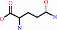 L-glutamine
|
+
|
H2O
|
=
|
 L-glutamate
|
+
|
NH4(+)
|
|
|
|
|
|
|
|
|
|
Enzyme class 3:
|
|
E.C.4.3.3.6
- pyridoxal 5'-phosphate synthase (glutamine hydrolyzing).
|
|
|
|
|
|
|
Reaction:
|
|
aldehydo-D-ribose 5-phosphate + D-glyceraldehyde 3-phosphate + L-glutamine = pyridoxal 5'-phosphate + L-glutamate + phosphate + 3 H2O + H+
|
|
|
|
|
|
aldehydo-D-ribose 5-phosphate
|
+
|
 D-glyceraldehyde 3-phosphate
|
+
|
L-glutamine
|
=
|
 pyridoxal 5'-phosphate
|
+
|
L-glutamate
|
+
|
 phosphate
|
+
|
3
×
H2O
|
+
|
H(+)
|
|
|
|
|
|
|
|
|
|
|
|
|
Note, where more than one E.C. class is given (as above), each may
correspond to a different protein domain or, in the case of polyprotein
precursors, to a different mature protein.
|
|
|
|
Molecule diagrams generated from .mol files obtained from the
KEGG ftp site
|
|
|
|
|
|
|
|
|
|
|
|
|
|
 |
|
|
|
|
|
|
| |
|
|
| |
|
DOI no:
|
J Biol Chem
281:3633-3641
(2006)
|
|
PubMed id:
|
|
|
|
|
|
| |
|
Vitamin B6 Biosynthesis by the Malaria Parasite Plasmodium falciparum: BIOCHEMICAL AND STRUCTURAL INSIGHTS.
|
|
M.Gengenbacher,
T.B.Fitzpatrick,
T.Raschle,
K.Flicker,
I.Sinning,
S.Müller,
P.Macheroux,
I.Tews,
B.Kappes.
|
|
|
|
|
| |
ABSTRACT
|
|
|
|
| |
|
|
Vitamin B6 is one of nature's most versatile cofactors. Most organisms
synthesize vitamin B6 via a recently discovered pathway employing the proteins
Pdx1 and Pdx2. Here we present an in-depth characterization of the respective
orthologs from the malaria parasite, Plasmodium falciparum. Expression profiling
of Pdx1 and -2 shows that blood-stage parasites indeed possess a functional
vitamin B6 de novo biosynthesis. Recombinant Pdx1 and Pdx2 form a complex that
functions as a glutamine amidotransferase with Pdx2 as the glutaminase and Pdx1
as pyridoxal-5 '-phosphate synthase domain. Complex formation is required for
catalytic activity of either domain. Pdx1 forms a chimeric bi-enzyme with the
bacterial YaaE, a Pdx2 ortholog, both in vivo and in vitro, although this
chimera does not attain full catalytic activity, emphasizing that
species-specific structural features govern the interaction between the protein
partners of the PLP synthase complexes in different organisms. To gain insight
into the activation mechanism of the parasite bi-enzyme complex, the
three-dimensional structure of Pdx2 was determined at 1.62 A. The obstruction of
the oxyanion hole indicates that Pdx2 is in a resting state and that activation
occurs upon Pdx1-Pdx2 complex formation.
|
|
|
|
|
|
| |
Selected figure(s)
|
|
|
|
| |
|
|

|
|

|
|
Figure 4.
Structural analysis of Pdx2. A, ribbon representation of the
x-ray structure of Pdx2. Amino acids Cys^87, His^196, and
Glu^198 make up the catalytic triad. The respective residues are
shown in yellow in all panels. Residues involved in the putative
interface with the synthase subunit are labeled in green.
Differences to the Yaa E ortholog are shown in red. B, stick
representation of the active site of Pdx2. The loop carrying the
nucleophilic cysteine comprises residues Gly^85, Thr^86, Cys^87,
Ala^88, and Gly^89. This loop is shown together with the 2F[o]
-F[c] electron density at a level of 1.2σ. The double
conformation of Cys^87 is visible. The proposed binding site of
the synthase subunit is indicated. C, the proposed oxyanion
hole, which forms during catalysis, is obstructed in Pdx2 by the
carbonyl of Gly^51. D, the oxyanion hole is formed in the
apo-form of CPS (1JDB) by the peptide nitrogens of Gly^241 and
Leu^270. E, in the glutamine-bound state of CPS (1A9X), this
conformation is maintained. The figure was prepared with PyMOL
(53).
|
|
Figure 5.
Cross-species interaction between Pdx1 and YaaE. A, growth of
the B. subtilis 168 (trpC2) YaaD disruptant complemented with
control construct lacking the ribosomal binding site (Pdx1) (1)
or the complementation construct (RBS-Pdx1) (2) on minimal
plates (TMM) without and with additives (0.05 mm pyridoxal
(TMM+pyridoxal) or 2% xylose (TMM+xylose)) in the presence or
absence of IPTG. IPTG is required to induce the expression of
the endogenous YaaE. B, growth curves of the B. subtilis 168
(trpC2) YaaD disruptant complemented with RBS-Pdx1 (filled
symbols: •, ▾, and ▪) or the RBS-lacking Pdx1 control
construct (open symbols: ○,▾, and □) in TMM plus IPTG
without and with additives (0.05 mm pyridoxal or 2% xylose).
•, RBS-Pdx1 in TMM; ○, Pdx1 in TMM; ○, RBS-Pdx1 in TMM
plus 0.05 mm pyridoxal;▵, Pdx1 in TMM plus 0.05 mm mm
pyridoxal; •, RBS-Pdx1 in TMM -Pdx1 expression was induced by
the addition of 2% xylose; □, Pdx1 in TMM plus 2% xylose. C,
PLP formation by the Pdx1-YaaE complex in the presence of
ribulose 5-phosphate, G3P, and 10 mm Gln: ▴, YaaD-YaaE complex
(1:1); •, Pdx1-Pdx2 complex (1:1); •, Pdx1-YaaE (1:5); ○,
Pdx1-YaaE (1:1);▵, YaaD; □, Pdx1; ♦, no enzyme.
|
|
|
|
|
|
| |
The above figures are
reprinted
by permission from the ASBMB:
J Biol Chem
(2006,
281,
3633-3641)
copyright 2006.
|
|
| |
Figures were
selected
by an automated process.
|
|
|
|
|
|
|
|
|
|
|
|
|
|
|
|
 |
 |
 |
|
Literature references that cite this PDB file's key reference
|
|
|
| |
PubMed id
|
|
Reference
|
|
|
|
|
|
T.B.Fitzpatrick,
C.Moccand,
and
C.Roux
(2010).
Vitamin B6 biosynthesis: charting the mechanistic landscape.
|
| |
Chembiochem,
11,
1185-1193.
|
|
|
|
|
|
|
T.Dick,
U.Manjunatha,
B.Kappes,
and
M.Gengenbacher
(2010).
Vitamin B6 biosynthesis is essential for survival and virulence of Mycobacterium tuberculosis.
|
| |
Mol Microbiol,
78,
980-988.
|
|
|
|
|
|
|
H.Chen,
and
L.Xiong
(2009).
The short-rooted vitamin B(6)-deficient mutant pdx1 has impaired local auxin biosynthesis.
|
| |
Planta,
229,
1303-1310.
|
|
|
|
|
|
|
H.Chen,
and
L.Xiong
(2009).
Enhancement of vitamin B(6) levels in seeds through metabolic engineering.
|
| |
Plant Biotechnol J,
7,
673-681.
|
|
|
|
|
|
|
I.B.Müller,
F.Wu,
B.Bergmann,
J.Knöckel,
R.D.Walter,
H.Gehring,
and
C.Wrenger
(2009).
Poisoning pyridoxal 5-phosphate-dependent enzymes: a new strategy to target the malaria parasite Plasmodium falciparum.
|
| |
PLoS ONE,
4,
e4406.
|
|
|
|
|
|
|
K.Benabdellah,
C.Azcón-Aguilar,
A.Valderas,
D.Speziga,
T.B.Fitzpatrick,
and
N.Ferrol
(2009).
GintPDX1 encodes a protein involved in vitamin B6 biosynthesis that is up-regulated by oxidative stress in the arbuscular mycorrhizal fungus Glomus intraradices.
|
| |
New Phytol,
184,
682-693.
|
|
|
|
|
|
|
T.Raschle,
D.Speziga,
W.Kress,
C.Moccand,
P.Gehrig,
N.Amrhein,
E.Weber-Ban,
and
T.B.Fitzpatrick
(2009).
Intersubunit cross-talk in pyridoxal 5'-phosphate synthase, coordinated by the C terminus of the synthase subunit.
|
| |
J Biol Chem,
284,
7706-7718.
|
|
|
|
|
|
|
I.B.Müller,
J.Knöckel,
M.R.Groves,
R.Jordanova,
S.E.Ealick,
R.D.Walter,
and
C.Wrenger
(2008).
The assembly of the plasmodial PLP synthase complex follows a defined course.
|
| |
PLoS ONE,
3,
e1815.
|
|
|
|
|
|
|
D.E.Scott,
A.Ciulli,
and
C.Abell
(2007).
Coenzyme biosynthesis: enzyme mechanism, structure and inhibition.
|
| |
Nat Prod Rep,
24,
1009-1026.
|
|
|
|
|
|
|
M.E.Webb,
A.Marquet,
R.R.Mendel,
F.Rébeillé,
and
A.G.Smith
(2007).
Elucidating biosynthetic pathways for vitamins and cofactors.
|
| |
Nat Prod Rep,
24,
988.
|
|
|
|
|
|
|
P.Gayathri,
H.Balaram,
and
M.R.Murthy
(2007).
Structural biology of plasmodial proteins.
|
| |
Curr Opin Struct Biol,
17,
744-754.
|
|
|
|
|
|
|
S.Müller,
and
B.Kappes
(2007).
Vitamin and cofactor biosynthesis pathways in Plasmodium and other apicomplexan parasites.
|
| |
Trends Parasitol,
23,
112-121.
|
|
|
|
|
|
|
S.Mouilleron,
and
B.Golinelli-Pimpaneau
(2007).
Conformational changes in ammonia-channeling glutamine amidotransferases.
|
| |
Curr Opin Struct Biol,
17,
653-664.
|
|
|
|
|
|
|
F.Zein,
Y.Zhang,
Y.N.Kang,
K.Burns,
T.P.Begley,
and
S.E.Ealick
(2006).
Structural insights into the mechanism of the PLP synthase holoenzyme from Thermotoga maritima.
|
| |
Biochemistry,
45,
14609-14620.
|
|
|
PDB code:
|
|
|
|
|
|
|
|
M.L.Eschbach,
I.B.Müller,
T.W.Gilberger,
R.D.Walter,
and
C.Wrenger
(2006).
The human malaria parasite Plasmodium falciparum expresses an atypical N-terminally extended pyrophosphokinase with specificity for thiamine.
|
| |
Biol Chem,
387,
1583-1591.
|
|
|
|
|
|
|
M.Strohmeier,
T.Raschle,
J.Mazurkiewicz,
K.Rippe,
I.Sinning,
T.B.Fitzpatrick,
and
I.Tews
(2006).
Structure of a bacterial pyridoxal 5'-phosphate synthase complex.
|
| |
Proc Natl Acad Sci U S A,
103,
19284-19289.
|
|
|
PDB codes:
|
|
|
|
|
|
|
|
O.Titiz,
M.Tambasco-Studart,
E.Warzych,
K.Apel,
N.Amrhein,
C.Laloi,
and
T.B.Fitzpatrick
(2006).
PDX1 is essential for vitamin B6 biosynthesis, development and stress tolerance in Arabidopsis.
|
| |
Plant J,
48,
933-946.
|
|
|
|
|
|
The most recent references are shown first.
Citation data come partly from CiteXplore and partly
from an automated harvesting procedure. Note that this is likely to be
only a partial list as not all journals are covered by
either method. However, we are continually building up the citation data
so more and more references will be included with time.
Where a reference describes a PDB structure, the PDB
code is
shown on the right.
|
|


 Links
Links


