 |
PDBsum entry 3fqf

|
|
|
|
|
|

|

|

|

|
|

|
|

|
|

|
|

|
|

|
|
|
 |

|

|
|
|
|
|
|
|
|
|
|
|
|
|
|
|
|
|
|
|
|
|
|
|
|
|
|
|
|
|
|
|
|
|
|
Oxidoreductase
|
PDB id
|
|
|
|
3fqf
|
|
|
|
|
|
|
|
|
|
|
|
|
|
|

|

|

|
|
|
|
|
|
|
|
 |
Contents |
 |
|
|
|
|
|
|
|
|
|
|
|
|
* Residue conservation analysis
|
|
|
|
|
|
|
|
|
|
|
Enzyme class:
|
|
E.C.1.5.1.3
- dihydrofolate reductase.
|
|
|
|
|
|
|
Pathway:
|
|
Folate Coenzymes
|
|
|
|
|
|
Reaction:
|
|
(6S)-5,6,7,8-tetrahydrofolate + NADP+ = 7,8-dihydrofolate + NADPH + H+
|
|
|
|
|
|
(6S)-5,6,7,8-tetrahydrofolate
|
+
|
NADP(+)
Bound ligand (Het Group name = )
corresponds exactly
|
=
|
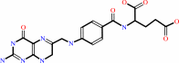 7,8-dihydrofolate
|
+
|
 NADPH
NADPH
|
+
|
H(+)
|
|
|
|
|
|
|
|
|
|
|
|
|
Molecule diagrams generated from .mol files obtained from the
KEGG ftp site
|
|
|
|
|
|
|
|
|
|
|
|
|
|
 |
|
|
|
|
|
|
| |
|
|
| |
|
DOI no:
|
J Mol Biol
387:1298-1308
(2009)
|
|
PubMed id:
|
|
|
|
|
|
| |
|
Crystal structures of wild-type and mutant methicillin-resistant Staphylococcus aureus dihydrofolate reductase reveal an alternate conformation of NADPH that may be linked to trimethoprim resistance.
|
|
K.M.Frey,
J.Liu,
M.N.Lombardo,
D.B.Bolstad,
D.L.Wright,
A.C.Anderson.
|
|
|
|
|
| |
ABSTRACT
|
|
|
|
| |
|
|
Both hospital- and community-acquired Staphylococcus aureus infections have
become major health concerns in terms of morbidity, suffering and cost.
Trimethoprim-sulfamethoxazole (TMP-SMZ) is an alternative treatment for
methicillin-resistant S. aureus (MRSA) infections. However, TMP-resistant
strains have arisen with point mutations in dihydrofolate reductase (DHFR), the
target for TMP. A single point mutation, F98Y, has been shown biochemically to
confer the majority of this resistance to TMP. Using a structure-based approach,
we have designed a series of novel propargyl-linked DHFR inhibitors that are
active against several trimethoprim-resistant enzymes. We screened this series
against wild-type and mutant (F98Y) S. aureus DHFR and found that several are
active against both enzymes and specifically that the meta-biphenyl class of
these inhibitors is the most potent. In order to understand the structural basis
of this potency, we determined eight high-resolution crystal structures: four
each of the wild-type and mutant DHFR enzymes bound to various propargyl-linked
DHFR inhibitors. In addition to explaining the structure-activity relationships,
several of the structures reveal a novel conformation for the cofactor, NADPH.
In this new conformation that is predominantly associated with the mutant
enzyme, the nicotinamide ring is displaced from its conserved location and three
water molecules complete a network of hydrogen bonds between the nicotinamide
ring and the protein. In this new position, NADPH has reduced interactions with
the inhibitor. An equilibrium between the two conformations of NADPH, implied by
their occupancies in the eight crystal structures, is influenced both by the
ligand and the F98Y mutation. The mutation induced equilibrium between two
NADPH-binding conformations may contribute to decrease TMP binding and thus may
be responsible for TMP resistance.
|
|
|
|
|
|
| |
Selected figure(s)
|
|
|
|
| |
|
|

|
|

|
|
Figure 1.
Fig. 1. Stereoview images of the wild-type (teal) and
Sa(F98Y) mutant (gold) enzymes bound to: a, compound 5
(wild-type light green, F98Y cyan); b, compound 8 (wild-type
orange, F98Y lavender); c, compound 10 (wild-type purple, F98Y
dark green); and d, compound 15 (wild-type yellow, F98Y gray).
The standard conformation of NADPH is shown in magenta and the
alternative conformation is shown in blue. Water molecules
(shown in Fig. 3) are omitted for clarity.
|
|
Figure 2.
Fig. 2. Electron density at the active site for
SaDHFR(F98Y):NADPH:5. Protein is shown with 2F[o]–F[c] density
(1.5 σ, blue) and ligands are shown with omit F[o]–F[c]
density (3.0 σ, grey). A full view of the density for the
alternative conformation is shown at the right.
|
|
|
|
|
|
| |
The above figures are
reprinted
by permission from Elsevier:
J Mol Biol
(2009,
387,
1298-1308)
copyright 2009.
|
|
| |
Figures were
selected
by the author.
|
|
|
|
|
|
|
|
|
|
|
|
|
|
|
|
 |
 |
 |
|
Literature references that cite this PDB file's key reference
|
|
|
| |
PubMed id
|
|
Reference
|
|
|
|
|
|
C.R.Bourne,
E.W.Barrow,
R.A.Bunce,
P.C.Bourne,
K.D.Berlin,
and
W.W.Barrow
(2010).
Inhibition of antibiotic-resistant Staphylococcus aureus by the broad-spectrum dihydrofolate reductase inhibitor RAB1.
|
| |
Antimicrob Agents Chemother,
54,
3825-3833.
|
|
|
PDB codes:
|
|
|
|
|
|
|
|
K.M.Frey,
I.Georgiev,
B.R.Donald,
and
A.C.Anderson
(2010).
Predicting resistance mutations using protein design algorithms.
|
| |
Proc Natl Acad Sci U S A,
107,
13707-13712.
|
|
|
PDB codes:
|
|
|
|
|
|
|
|
K.M.Frey,
M.N.Lombardo,
D.L.Wright,
and
A.C.Anderson
(2010).
Towards the understanding of resistance mechanisms in clinically isolated trimethoprim-resistant, methicillin-resistant Staphylococcus aureus dihydrofolate reductase.
|
| |
J Struct Biol,
170,
93-97.
|
|
|
PDB code:
|
|
|
|
|
|
|
The most recent references are shown first.
Citation data come partly from CiteXplore and partly
from an automated harvesting procedure. Note that this is likely to be
only a partial list as not all journals are covered by
either method. However, we are continually building up the citation data
so more and more references will be included with time.
Where a reference describes a PDB structure, the PDB
codes are
shown on the right.
|
|


 Links
Links


