 |
PDBsum entry 2vyc

|
|
|
|
|
 |
Contents |
 |
|
|
|
|
|
|
|
|
|
|
|
|
* Residue conservation analysis
|
|
|
|
|
|
PDB id:
|
|
|
|
| Name: |
|
Lyase
|
|
|
Title:
|
|
Crystal structure of acid induced arginine decarboxylase from e. Coli
|
|
Structure:
|
|
Biodegradative arginine decarboxylase. Chain: a, b, c, d, e, f, g, h, i, j. Synonym: arginine decarboxylase. Other_details: covelent bond between cofactor pyridoxal 5' phosphate (plp) and residue lys386.
|
|
Source:
|
|
Escherichia coli. Organism_taxid: 469008. Strain: bl21(de3)
|
|
Resolution:
|
|
|
2.40Å
|
R-factor:
|
0.179
|
R-free:
|
0.229
|
|
|
Authors:
|
|
J.Andrell,M.G.Hicks,T.Palmer,E.P.Carpenter,S.Iwata,M.J.Maher
|
|
Key ref:
|
|
J.Andréll
et al.
(2009).
Crystal structure of the acid-induced arginine decarboxylase from Escherichia coli: reversible decamer assembly controls enzyme activity.
Biochemistry,
48,
3915-3927.
PubMed id:
|
|
|
Date:
|
|
|
22-Jul-08
|
Release date:
|
31-Mar-09
|
|
|
|
|
|
|
PROCHECK
|

|
|
|
|
|
Headers
|
|
|
|
References
|
|
|
|
|
|

|
|
P28629
(ADIA_ECOLI) -
Biodegradative arginine decarboxylase from Escherichia coli (strain K12)
|
|
|
|
Seq:
Struc:
|
|
|
|
755 a.a.
755 a.a.
|
|

|

|
|
|
|
|
|
|
|
|
|
|
|
Key: |
 |
PfamA domain |
|
|
 |
Secondary structure |
|
 |
CATH domain |
|
|
|
|
|
|
|
|
|
|
|
|
|
Enzyme class:
|
|
E.C.4.1.1.19
- arginine decarboxylase.
|
|
|
|
|
|
|
Reaction:
|
|
L-arginine + H+ = agmatine + CO2
|
|
|
|
|
|
 L-arginine
|
+
|
H(+)
|
=
|
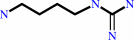 agmatine
|
+
|
 CO2
CO2
|
|
|
|
|
|
|
|
|
|
Cofactor:
|
|
Pyridoxal 5'-phosphate
|
|
|
|
|
|
Pyridoxal 5'-phosphate
Bound ligand (Het Group name =
PLP)
matches with 93.75% similarity
|
|
|
|
|
|
|
Molecule diagrams generated from .mol files obtained from the
KEGG ftp site
|
|
|
|
|
|
|
|
|
|
|
|
|
|
 |
|
|
|
|
|
|
| |
|
|
| |
|
|
Biochemistry
48:3915-3927
(2009)
|
|
PubMed id:
|
|
|
|
|
|
| |
|
Crystal structure of the acid-induced arginine decarboxylase from Escherichia coli: reversible decamer assembly controls enzyme activity.
|
|
J.Andréll,
M.G.Hicks,
T.Palmer,
E.P.Carpenter,
S.Iwata,
M.J.Maher.
|
|
|
|
|
| |
ABSTRACT
|
|
|
|
| |
|
|
The acid-induced arginine decarboxylase is part of an enzymatic system in
Escherichia coli that contributes to making this organism acid resistant. The
arginine decarboxylase is a vitamin B(6)-dependent enzyme that is active at
acidic pH. It consumes a proton in the decarboxylation of arginine to agmatine,
and by working in tandem with an arginine-agmatine antiporter, this enzymatic
cycle protects the organism by preventing the accumulation of protons inside the
cell. We have determined the structure of the acid-induced arginine
decarboxylase by X-ray crystallography to 2.4 A resolution. The arginine
decarboxylase structure revealed a ca. 800 kDa decamer composed as a pentamer of
five homodimers. Each homodimer has an abundance of acidic surface residues,
which at neutral pH prevents inactive homodimers from associating into active
decamers. Conversely, acidic conditions favor the assembly of active decamers.
Therefore, the structure of arginine decarboxylase presents a mechanism by which
its activity is modulated by external pH.
|
|
|
|
|
|
|
|
|
|
|
|
|
|
|
|
|
|
 |
 |
 |
|
Literature references that cite this PDB file's key reference
|
|
|
| |
PubMed id
|
|
Reference
|
|
|
|
|
|
U.Kanjee,
I.Gutsche,
E.Alexopoulos,
B.Zhao,
M.El Bakkouri,
G.Thibault,
K.Liu,
S.Ramachandran,
J.Snider,
E.F.Pai,
and
W.A.Houry
(2011).
Linkage between the bacterial acid stress and stringent responses: the structure of the inducible lysine decarboxylase.
|
| |
EMBO J,
30,
931-944.
|
|
|
PDB codes:
|
|
|
|
|
|
|
|
B.Zhao,
and
W.A.Houry
(2010).
Acid stress response in enteropathogenic gammaproteobacteria: an aptitude for survival.
|
| |
Biochem Cell Biol,
88,
301-314.
|
|
|
|
|
|
|
F.Karbassi,
V.Quiros,
V.Pancholi,
and
M.J.Kornblatt
(2010).
Dissociation of the octameric enolase from S. pyogenes--one interface stabilizes another.
|
| |
PLoS One,
5,
e8810.
|
|
|
|
|
|
The most recent references are shown first.
Citation data come partly from CiteXplore and partly
from an automated harvesting procedure. Note that this is likely to be
only a partial list as not all journals are covered by
either method. However, we are continually building up the citation data
so more and more references will be included with time.
Where a reference describes a PDB structure, the PDB
codes are
shown on the right.
|
|


 Links
Links


