 |
PDBsum entry 1bkp

|
|
|
|
|
|

|

|

|

|
|

|
|

|
|

|
|

|
|
|
 |

|

|
|
|
|
|
|
|
|
|
|
|
|
|
|
|
|
|
|
|
|
|
|
|
|
|
|
|
|
|
|
|
|
|
Methyltransferase
|
PDB id
|
|
|
|
1bkp
|
|
|
|
|
|
|
|
|
|
|
|
|
|
|

|

|

|
|
|
|
|
|
|
|
 |
Contents |
 |
|
|
|
|
|
|
|
|
|
|
* Residue conservation analysis
|
|
|
|
|
|
|
|
|
|
|
Enzyme class:
|
|
E.C.2.1.1.45
- thymidylate synthase.
|
|
|
|
|
|
|
Pathway:
|
|
Folate Coenzymes
|
|
|
|
|
|
Reaction:
|
|
dUMP + (6R)-5,10-methylene-5,6,7,8-tetrahydrofolate = 7,8-dihydrofolate + dTMP
|
|
|
|
|
|
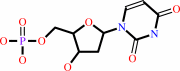 dUMP
dUMP
|
+
|
(6R)-5,10-methylene-5,6,7,8-tetrahydrofolate
|
=
|
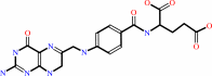 7,8-dihydrofolate
|
+
|
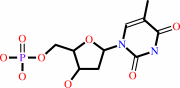 dTMP
dTMP
|
|
|
|
|
|
|
|
|
|
|
|
|
Molecule diagrams generated from .mol files obtained from the
KEGG ftp site
|
|
|
|
|
|
|
|
|
|
|
|
|
|
 |
|
|
|
|
|
|
| |
|
|
| |
|
DOI no:
|
Biochemistry
37:14736-14747
(1998)
|
|
PubMed id:
|
|
|
|
|
|
| |
|
Crystal structures of a unique thermal-stable thymidylate synthase from Bacillus subtilis.
|
|
T.J.Stout,
U.Schellenberger,
D.V.Santi,
R.M.Stroud.
|
|
|
|
|
| |
ABSTRACT
|
|
|
|
| |
|
|
Unlike all other organisms studied to date, Bacillus subtilis expresses two
different thymidylate synthases: bsTS-A and bsTS-B. bsTS-A displays enhanced
enzymatic and structural thermal stability uncharacteristic of most TSs. Despite
the high level of TS conservation across most species, bsTS-A shares low
sequence identity (<40%) with the majority of TSs from other organisms. This
TS and the TSs from Lactococcus lactis and phage Phi3T-to which it is most
similar-have been of interest for some time since, by structure-based sequence
alignment, they appear to lack several key residues shown by mutagenesis to be
essential to enzymatic function [Greene, P. J., Yu, P. L., Zhao, J., Schiffer,
C. A., and Santi, D. (1994) Protein Sci. 3, 1114-6]. In addition, bsTS-A
demonstrates specific activity 2-3-fold higher than TS from Lactobacillus casei
or Escherichia coli. We have solved the crystal structure of this unusual TS in
four crystal forms to a maximum resolution of 1.7 A. Each of these crystal forms
contains either one or two noncrystallographically related dimers. Stabilization
of the beta-sheet dimer interface through a dramatic architecture of buttressed
internal salt bridges maintains the structural integrity of bsTS-A at elevated
temperatures. Melting curves of TSs from L. casei and E. coli are compared to
that of TS-A from B. subtilis and correlated with numbers of hydrogen bonds,
salt bridges, and the numbers of interactions localized to the dimer interface.
Analysis of this structure will shed light on the conservation of function
across diversity of sequence, as well as provide insights into the thermal
stabilization of a highly conserved enzyme.
|
|
|
|
|
|
|
|
|
|
|
|
|
|
|
|
|
|
 |
 |
 |
|
Literature references that cite this PDB file's key reference
|
|
|
| |
PubMed id
|
|
Reference
|
|
|
|
|
|
R.Almog,
C.A.Waddling,
F.Maley,
G.F.Maley,
and
P.Van Roey
(2001).
Crystal structure of a deletion mutant of human thymidylate synthase Delta (7-29) and its ternary complex with Tomudex and dUMP.
|
| |
Protein Sci,
10,
988-996.
|
|
|
PDB codes:
|
|
|
|
|
|
|
|
H.K.Song,
S.H.Sohn,
and
S.W.Suh
(1999).
Crystal structure of deoxycytidylate hydroxymethylase from bacteriophage T4, a component of the deoxyribonucleoside triphosphate-synthesizing complex.
|
| |
EMBO J,
18,
1104-1113.
|
|
|
PDB codes:
|
|
|
|
|
|
|
|
K.M.Fox,
F.Maley,
A.Garibian,
L.M.Changchien,
and
P.Van Roey
(1999).
Crystal structure of thymidylate synthase A from Bacillus subtilis.
|
| |
Protein Sci,
8,
538-544.
|
|
|
PDB code:
|
|
|
|
|
|
|
The most recent references are shown first.
Citation data come partly from CiteXplore and partly
from an automated harvesting procedure. Note that this is likely to be
only a partial list as not all journals are covered by
either method. However, we are continually building up the citation data
so more and more references will be included with time.
Where a reference describes a PDB structure, the PDB
codes are
shown on the right.
|
|


 Links
Links


