 |
PDBsum entry 3ocn

|
|
|
|
|
|

|

|

|

|
|

|
|

|
|

|
|

|
|

|
|
|
 |

|

|
|
|
|
|
|
|
|
|
|
|
|
|
|
|
|
|
|
|
|
|
|
|
|
|
|
|
|
|
|
|
|
|
|
Penicillin-binding protein/antibiotic
|
PDB id
|
|
|
|
3ocn
|
|
|
|
|
|
|
|
|
|
|
|
|
|
|

|

|

|
|
|
|
|
|
|
|
 |
Contents |
 |
|
|
|
|
|
|
|
|
|
|
|
|
* Residue conservation analysis
|
|
|
|
|
|
|
|
|
|
|
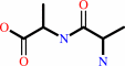
Enzyme class:
|
|
E.C.3.4.16.4
- serine-type D-Ala-D-Ala carboxypeptidase.
|
|
|
|
|
|
|
Reaction:
|
|
D-alanyl-D-alanine + H2O = 2 D-alanine
|
|
|
|
|
|
|
+
|
|
=
|
 2
×
2
×
|
|
|
|
|
|
|
|
|
|
|
|
|
Molecule diagrams generated from .mol files obtained from the
KEGG ftp site
|
|
|
|
|
|
|
|
|
|
|
|
|
|
 |
|
|
|
|
|
|
| |
|
|
| |
|
|
J Mol Biol
405:173-184
(2011)
|
|
PubMed id:
|
|
|
|
|
|
| |
|
Crystal structures of penicillin-binding protein 3 from Pseudomonas aeruginosa: comparison of native and antibiotic-bound forms.
|
|
S.Sainsbury,
L.Bird,
V.Rao,
S.M.Shepherd,
D.I.Stuart,
W.N.Hunter,
R.J.Owens,
J.Ren.
|
|
|
|
|
| |
ABSTRACT
|
|
|
|
| |
|
|
We report the first crystal structures of a penicillin-binding protein (PBP),
PBP3, from Pseudomonas aeruginosa in native form and covalently linked to two
important β-lactam antibiotics, carbenicillin and ceftazidime. Overall, the
structures of apo and acyl complexes are very similar; however, variations in
the orientation of the amino-terminal membrane-proximal domain relative to that
of the carboxy-terminal transpeptidase domain indicate interdomain flexibility.
Binding of either carbenicillin or ceftazidime to purified PBP3 increases the
thermostability of the enzyme significantly and is associated with local
conformational changes, which lead to a narrowing of the substrate-binding
cleft. The orientations of the two β-lactams in the active site and the key
interactions formed between the ligands and PBP3 are similar despite differences
in the two drugs, indicating a degree of flexibility in the binding site. The
conserved binding mode of β-lactam-based inhibitors appears to extend to other
PBPs, as suggested by a comparison of the PBP3/ceftazidime complex and the
Escherichia coli PBP1b/ceftoxamine complex. Since P. aeruginosa is an important
human pathogen, the structural data reveal the mode of action of the frontline
antibiotic ceftazidime at the molecular level. Improved drugs to combat
infections by P. aeruginosa and related Gram-negative bacteria are sought and
our study provides templates to assist that process and allows us to discuss new
ways of inhibiting PBPs.
|
|
|
|
|
|
|
|
|
|
|
|
|
|
|
|
|
|
 |
 |
 |
|


 Links
Links


