 |
PDBsum entry 1ia4

|
|
|
|
|
|

|

|

|

|
|

|
|

|
|

|
|

|
|

|
|

|
|
|
 |

|

|
|
|
|
|
|
|
|
|
|
|
|
|
|
|
|
|
|
|
|
|
|
|
|
|
|
|
|
|
|
|
|
|
|
|
Oxidoreductase
|
PDB id
|
|
|
|
1ia4
|
|
|
|
|
|
|
|
|
|
|
|
|
|
|

|

|

|
|
|
|
|
|
|
|
 |
Contents |
 |
|
|
|
|
|
|
|
|
|
|
|
|
* Residue conservation analysis
|
|
|
|
|
|
|
|
|
|
|
Enzyme class:
|
|
E.C.1.5.1.3
- dihydrofolate reductase.
|
|
|
|
|
|
|
Pathway:
|
|
Folate Coenzymes
|
|
|
|
|
|
Reaction:
|
|
(6S)-5,6,7,8-tetrahydrofolate + NADP+ = 7,8-dihydrofolate + NADPH + H+
|
|
|
|
|
|
(6S)-5,6,7,8-tetrahydrofolate
|
+
|
NADP(+)
Bound ligand (Het Group name = )
matches with 72.92% similarity
|
=
|
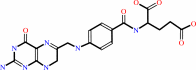 7,8-dihydrofolate
|
+
|
 NADPH
NADPH
|
+
|
H(+)
|
|
|
|
|
|
|
|
|
|
|
|
|
Molecule diagrams generated from .mol files obtained from the
KEGG ftp site
|
|
|
|
|
|
|
|
|
|
|
|
|
|
 |
|
|
|
|
|
|
| |
|
|
| |
|
DOI no:
|
J Med Chem
44:2928-2932
(2001)
|
|
PubMed id:
|
|
|
|
|
|
| |
|
X-Ray crystal structures of Candida albicans dihydrofolate reductase: high resolution ternary complexes in which the dihydronicotinamide moiety of NADPH is displaced by an inhibitor.
|
|
M.Whitlow,
A.J.Howard,
D.Stewart,
K.D.Hardman,
J.H.Chan,
D.P.Baccanari,
R.L.Tansik,
J.S.Hong,
L.F.Kuyper.
|
|
|
|
|
| |
ABSTRACT
|
|
|
|
| |
|
|
X-ray crystallographic analysis of 5-(4'-substituted
phenyl)sulfanyl-2,4-diaminoquinazoline inhibitors in ternary complex with
Candida albicans dihydrofolate reductase (DHFR) and NADPH revealed two distinct
modes of binding. The two compounds with small 4'-substituents (H and CH3) were
found to bind with the phenyl group oriented in the plane of the quinazoline
ring system and positioned adjacent to the C-helix. In contrast, the more
selective inhibitors with larger 4'-substituents (tert-butyl and N-morpholino)
were bound to the enzyme with the phenyl group perpendicular to the quinazoline
ring and positioned in the region of the active site that typically binds the
dihydronicotinamide moiety of NADPH. The cofactor appeared bound to DHFR but
with the disordered dihydronicotinamide swung away from the protein surface and
into solution. This unusual inhibitor binding mode may play an important role in
the high DHFR selectivity of these compounds and also may provide new ideas for
inhibitor design.
|
|
|
|
|
|
|
|
|
|
|
|
|
|
|
|
|
|
 |
 |
 |
|
Literature references that cite this PDB file's key reference
|
|
|
| |
PubMed id
|
|
Reference
|
|
|
|
|
|
C.R.Bourne,
R.A.Bunce,
P.C.Bourne,
K.D.Berlin,
E.W.Barrow,
and
W.W.Barrow
(2009).
Crystal structure of Bacillus anthracis dihydrofolate reductase with the dihydrophthalazine-based trimethoprim derivative RAB1 provides a structural explanation of potency and selectivity.
|
| |
Antimicrob Agents Chemother,
53,
3065-3073.
|
|
|
PDB codes:
|
|
|
|
|
|
|
|
J.L.Paulsen,
J.Liu,
D.B.Bolstad,
A.E.Smith,
N.D.Priestley,
D.L.Wright,
and
A.C.Anderson
(2009).
In vitro biological activity and structural analysis of 2,4-diamino-5-(2'-arylpropargyl)pyrimidine inhibitors of Candida albicans.
|
| |
Bioorg Med Chem,
17,
4866-4872.
|
|
|
|
|
|
|
J.Liu,
D.B.Bolstad,
A.E.Smith,
N.D.Priestley,
D.L.Wright,
and
A.C.Anderson
(2008).
Structure-guided development of efficacious antifungal agents targeting Candida glabrata dihydrofolate reductase.
|
| |
Chem Biol,
15,
990-996.
|
|
|
PDB code:
|
|
|
|
|
|
|
|
S.Henrich,
S.Richter,
and
R.C.Wade
(2008).
On the use of PIPSA to guide target-selective drug design.
|
| |
ChemMedChem,
3,
413-417.
|
|
|
|
|
|
|
K.H.Kim
(2007).
Outliers in SAR and QSAR: is unusual binding mode a possible source of outliers?
|
| |
J Comput Aided Mol Des,
21,
63-86.
|
|
|
|
|
|
|
M.Kontoyianni,
G.S.Sokol,
and
L.M.McClellan
(2005).
Evaluation of library ranking efficacy in virtual screening.
|
| |
J Comput Chem,
26,
11-22.
|
|
|
|
|
|
|
O.Senkovich,
V.Bhatia,
N.Garg,
and
D.Chattopadhyay
(2005).
Lipophilic antifolate trimetrexate is a potent inhibitor of Trypanosoma cruzi: prospect for chemotherapy of Chagas' disease.
|
| |
Antimicrob Agents Chemother,
49,
3234-3238.
|
|
|
|
|
|
|
R.Brenk,
J.J.Irwin,
and
B.K.Shoichet
(2005).
Here be dragons: docking and screening in an uncharted region of chemical space.
|
| |
J Biomol Screen,
10,
667-674.
|
|
|
|
|
|
|
R.Shi,
and
S.X.Lin
(2004).
Cofactor hydrogen bonding onto the protein main chain is conserved in the short chain dehydrogenase/reductase family and contributes to nicotinamide orientation.
|
| |
J Biol Chem,
279,
16778-16785.
|
|
|
PDB codes:
|
|
|
|
|
|
|
|
C.A.Bottoms,
P.E.Smith,
and
J.J.Tanner
(2002).
A structurally conserved water molecule in Rossmann dinucleotide-binding domains.
|
| |
Protein Sci,
11,
2125-2137.
|
|
|
|
|
|
The most recent references are shown first.
Citation data come partly from CiteXplore and partly
from an automated harvesting procedure. Note that this is likely to be
only a partial list as not all journals are covered by
either method. However, we are continually building up the citation data
so more and more references will be included with time.
Where a reference describes a PDB structure, the PDB
codes are
shown on the right.
|
|


 Links
Links


