Function and Biology

Crystal structure of 6-aminohexanoate-dimer hydrolase S112A/G181D/R187G/H266N/D370Y mutant complexd with 6-aminohexanoate

Source organism: Paenarthrobacter ureafaciens
Biochemical function: not assigned
Biological process: not assigned
Cellular component: not assigned

EC 3.5.1.46: 6-aminohexanoate-oligomer exohydrolase

Reaction catalysed:
(N-(6-aminohexanoyl))(n) + H(2)O = (N-(6-aminohexanoyl))(n-1) + 6-aminohexanoate
Systematic name:
N-(6-aminohexanoyl)-6-aminohexanoate exoamidohydrolase
Alternative Name(s):
  • 6-aminohexanoate-dimer hydrolase
  • 6-aminohexanoic acid oligomer hydrolase
  • N-(6-aminohexanoyl)-6-aminohexanoate amidohydrolase
  • NylB (gene name)
  • Nylon-6 hydrolase

GO terms

Biochemical function:
  • not assigned
Biological process:
  • not assigned
Cellular component:
  • not assigned

Sequence family

Pfam Protein family (Pfam)
PF00144
Domain description: Beta-lactamase
Occurring in:
  1. 6-aminohexanoate-dimer hydrolase
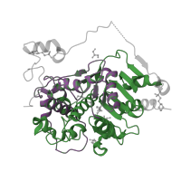
The deposited structure of PDB entry 3vwr contains 2 copies of Pfam domain PF00144 (Beta-lactamase) in 6-aminohexanoate-dimer hydrolase. Showing 2 copies in chain A.

InterPro InterPro annotations
IPR012338
Domain description: Beta-lactamase/transpeptidase-like
Occurring in:
  1. 6-aminohexanoate-dimer hydrolase
IPR001466
Domain description: Beta-lactamase-related
Occurring in:
  1. 6-aminohexanoate-dimer hydrolase

Structure domains

CATH CATH domains
3.40.710.10
Class: Alpha Beta
Architecture: 3-Layer(aba) Sandwich
Topology: Beta-lactamase
Homology: DD-peptidase/beta-lactamase superfamily
Occurring in:
  1. 6-aminohexanoate-dimer hydrolase
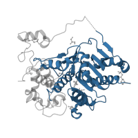
The deposited structure of PDB entry 3vwr contains 1 copy of CATH domain 3.40.710.10 (Beta-lactamase) in 6-aminohexanoate-dimer hydrolase. Showing 1 copy in chain A.
1.20.58.710
Class: Mainly Alpha
Architecture: Up-down Bundle
Topology: Methane Monooxygenase Hydroxylase; Chain G, domain 1
Homology: Methane Monooxygenase Hydroxylase; Chain G, domain 1
Occurring in:
  1. 6-aminohexanoate-dimer hydrolase
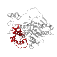
The deposited structure of PDB entry 3vwr contains 1 copy of CATH domain 1.20.58.710 (Methane Monooxygenase Hydroxylase; Chain G, domain 1) in 6-aminohexanoate-dimer hydrolase. Showing 1 copy in chain A.