 |
PDBsum entry 1s3y

|
|
|
|
|
|

|

|

|

|
|

|
|

|
|

|
|

|
|
|
 |

|

|
|
|
|
|
|
|
|
|
|
|
|
|
|
|
|
|
|
|
|
|
|
|
|
|
|
|
|
|
|
|
|
|
Oxidoreductase
|
PDB id
|
|
|
|
1s3y
|
|
|
|
|
|
|
|
|
|
|
|
|
|
|

|

|

|
|
|
|
|
|
|
|
 |
Contents |
 |
|
|
|
|
|
|
|
|
|
|
|
|
* Residue conservation analysis
|
|
|
|
|
|
|
|
|
|
|
Enzyme class:
|
|
E.C.1.5.1.3
- dihydrofolate reductase.
|
|
|
|
|
|
|
Pathway:
|
|
Folate Coenzymes
|
|
|
|
|
|
Reaction:
|
|
(6S)-5,6,7,8-tetrahydrofolate + NADP+ = 7,8-dihydrofolate + NADPH + H+
|
|
|
|
|
|
(6S)-5,6,7,8-tetrahydrofolate
|
+
|
NADP(+)
Bound ligand (Het Group name = )
corresponds exactly
|
=
|
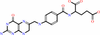 7,8-dihydrofolate
|
+
|
 NADPH
NADPH
|
+
|
H(+)
|
|
|
|
|
|
|
|
|
|
|
|
|
Molecule diagrams generated from .mol files obtained from the
KEGG ftp site
|
|
|
|
|
|
|
|
|
|
|
|
|
|
 |
|
|
|
|
|
|
| |
|
|
| |
|
DOI no:
|
Acta Crystallogr D Biol Crystallogr
60:646-655
(2004)
|
|
PubMed id:
|
|
|
|
|
|
| |
|
Structure determination of tetrahydroquinazoline antifolates in complex with human and Pneumocystis carinii dihydrofolate reductase: correlations between enzyme selectivity and stereochemistry.
|
|
V.Cody,
J.R.Luft,
W.Pangborn,
A.Gangjee,
S.F.Queener.
|
|
|
|
|
| |
ABSTRACT
|
|
|
|
| |
|
|
Structural data are reported for the first examples of the tetrahydroquinazoline
antifolate
(6R,6S)-2,4-diamino-6-(1-indolinomethyl)-5,6,7,8-tetrahydroquinazoline (1) and
its trimethoxy analogue
(6R,6S)-2,4-diamino-6-(3',4',5'-trimethoxybenzyl)-5,6,7,8-tetrahydroquinazoline
(2) as inhibitor complexes with dihydrofolate reductase (DHFR) from human
(hDHFR) and Pneumocystis carinii (pcDHFR) sources. The indoline analogue (1) was
crystallized as ternary complexes with NADPH and hDHFR (1.9 A resolution) and
pcDHFR (2.3 A resolution), while the trimethoxy quinazoline analogue (2) was
crystallized as a binary complex with hDHFR in two polymorphic rhombohedral R3
lattices: R3(1) to 1.8 A resolution and R3(2) to 2.0 A resolution. Structural
analysis of these potent and selective DHFR-inhibitor complexes revealed
preferential binding of the 6S-equatorial isomer in each structure. This
configuration is similar to that of the natural tetrahydrofolate substrate; that
is, 6S. These data also show that in both the hDHFR and pcDHFR ternary complexes
with (1) the indoline ring is partially disordered, with two static
conformations that differ between structures. These conformers also differ from
that observed for the trimethoxybenzyl ring of tetrahydroquinazoline (2). There
is also a correlation between the disorder of the flexible loop 23 and the
disorder of the cofactor nicotinamide ribose ring in the pcDHFR-NADPH-(1)
ternary complex. Comparison of the Toxoplasma gondii DHFR (tgDHFR) sequence with
those of other DHFRs provides insight into the role of sequence and conformation
in inhibitor-binding preferences which may aid in the design of novel
antifolates with specific DHFR selectivity.
|
|
|
|
|
|
| |
Selected figure(s)
|
|
|
|
| |
|
|

|
|

|
|
Figure 2.
Figure 2 Orientation of the pteridine ring for folates (cyan)
and antifolates (red). The Connolly surface for hDHFR is shown
in green and the contact distances of the nicotinamide ring of
NADPH are shown to the folate-reduction site.
|
|
Figure 8.
Figure 8 Comparison of the binding of inhibitor (1) to human
DHFR (violet) and pcDHFR (yellow). In each structure the
indoline ring of (1) is disordered. There are two orientations
observed in the human DHFR structure (violet) and two
orientations observed in the pcDHFR complex (green and cyan).
Diagram produced with SETOR (Evans, 1993[Evans, S. V. (1993). J.
Mol. Graph. 11, 134-138.]).
|
|
|
|
|
|
| |
The above figures are
reprinted
by permission from the IUCr:
Acta Crystallogr D Biol Crystallogr
(2004,
60,
646-655)
copyright 2004.
|
|
| |
Figures were
selected
by the author.
|
|
|
|
|
|
|
|
|
|
|
|
|
|
|
|
 |
 |
 |
|
Literature references that cite this PDB file's key reference
|
|
|
| |
PubMed id
|
|
Reference
|
|
|
|
|
|
M.Sáez-Ayala,
L.Sánchez-Del-Campo,
M.F.Montenegro,
S.Chazarra,
A.Tárraga,
J.Cabezas-Herrera,
and
J.N.Rodríguez-López
(2011).
Comparison of a pair of synthetic tea-catechin-derived epimers: synthesis, antifolate activity, and tyrosinase-mediated activation in melanoma.
|
| |
ChemMedChem,
6,
440-449.
|
|
|
|
|
|
|
J.P.Priestle
(2009).
3-D clustering: a tool for high throughput docking.
|
| |
J Mol Model,
15,
551-560.
|
|
|
|
|
|
|
J.P.Volpato,
B.J.Yachnin,
J.Blanchet,
V.Guerrero,
L.Poulin,
E.Fossati,
A.M.Berghuis,
and
J.N.Pelletier
(2009).
Multiple conformers in active site of human dihydrofolate reductase F31R/Q35E double mutant suggest structural basis for methotrexate resistance.
|
| |
J Biol Chem,
284,
20079-20089.
|
|
|
PDB code:
|
|
|
|
|
|
|
|
L.Sánchez-Del-Campo,
M.Sáez-Ayala,
S.Chazarra,
J.Cabezas-Herrera,
and
J.N.Rodríguez-López
(2009).
Binding of natural and synthetic polyphenols to human dihydrofolate reductase.
|
| |
Int J Mol Sci,
10,
5398-5410.
|
|
|
|
|
|
|
M.Spina,
M.Cuccioloni,
M.Mozzicafreddo,
F.Montecchia,
S.Pucciarelli,
A.M.Eleuteri,
E.Fioretti,
and
M.Angeletti
(2008).
Mechanism of inhibition of wt-dihydrofolate reductase from E. coli by tea epigallocatechin-gallate.
|
| |
Proteins,
72,
240-251.
|
|
|
|
|
|
|
N.Schormann,
O.Senkovich,
K.Walker,
D.L.Wright,
A.C.Anderson,
A.Rosowsky,
S.Ananthan,
B.Shinkre,
S.Velu,
and
D.Chattopadhyay
(2008).
Structure-based approach to pharmacophore identification, in silico screening, and three-dimensional quantitative structure-activity relationship studies for inhibitors of Trypanosoma cruzi dihydrofolate reductase function.
|
| |
Proteins,
73,
889-901.
|
|
|
PDB codes:
|
|
|
|
|
|
|
|
Z.Luka,
S.Pakhomova,
L.V.Loukachevitch,
M.Egli,
M.E.Newcomer,
and
C.Wagner
(2007).
5-methyltetrahydrofolate is bound in intersubunit areas of rat liver folate-binding protein glycine N-methyltransferase.
|
| |
J Biol Chem,
282,
4069-4075.
|
|
|
PDB codes:
|
|
|
|
|
|
|
|
G.A.Landrum,
J.E.Penzotti,
and
S.Putta
(2006).
Feature-map vectors: a new class of informative descriptors for computational drug discovery.
|
| |
J Comput Aided Mol Des,
20,
751-762.
|
|
|
|
|
|
|
V.Cody,
J.Pace,
K.Chisum,
and
A.Rosowsky
(2006).
New insights into DHFR interactions: analysis of Pneumocystis carinii and mouse DHFR complexes with NADPH and two highly potent 5-(omega-carboxy(alkyloxy) trimethoprim derivatives reveals conformational correlations with activity and novel parallel ring stacking interactions.
|
| |
Proteins,
65,
959-969.
|
|
|
PDB codes:
|
|
|
|
|
|
|
The most recent references are shown first.
Citation data come partly from CiteXplore and partly
from an automated harvesting procedure. Note that this is likely to be
only a partial list as not all journals are covered by
either method. However, we are continually building up the citation data
so more and more references will be included with time.
Where a reference describes a PDB structure, the PDB
code is
shown on the right.
|
|


 Links
Links


