 |
PDBsum entry 7nlp
|
|

|
|
PDB id:
|
|
|
|
| Name: |
|
Transferase
|
|
|
Title:
|
|
Crystal structure of mycobacterium tuberculosis argb in complex with l-canavanine
|
|
Structure:
|
|
Acetylglutamate kinase. Chain: a. Synonym: n-acetyl-l-glutamate 5-phosphotransferase,NAG kinase,nagk. Engineered: yes
|
|
Source:
|
|
Mycobacterium tuberculosis h37rv. Organism_taxid: 83332. Gene: argb, rv1654, mtcy06h11.19. Expressed in: escherichia coli bl21(de3). Expression_system_taxid: 469008
|
|
Resolution:
|
|
|
2.21Å
|
R-factor:
|
0.207
|
R-free:
|
0.241
|
|
|
Authors:
|
|
V.Mendes,S.E.Thomas,J.Cory-Wright,T.L.Blundell
|
|
Key ref:
|
|
P.Gupta
et al.
(2021).
A fragment-Based approach to assess the ligandability argb, Argc, Argd and argf in the l-Arginine biosynthe pathway of mycobacterium tuberculosis.
Comput struct biotechnol j,
19,
3491.
|
|
|
Date:
|
|
|
22-Feb-21
|
Release date:
|
30-Jun-21
|
|
|
|
|
|
|
PROCHECK
|

|
|
|
|
|
Headers
|
|
|
|
References
|
|
|
|
|
|

|
|
P9WQ01
(ARGB_MYCTU) -
Acetylglutamate kinase from Mycobacterium tuberculosis (strain ATCC 25618 / H37Rv)
|
|
|
|
Seq:
Struc:
|
|
|
|
294 a.a.
292 a.a.
|
|

|

|

|
|
|
|
|
|
|
|
|
|
|
Key: |
 |
PfamA domain |
|
|
 |
Secondary structure |
|
|
|
|
|
|
|
|
|
|
|
|
|
Enzyme class:
|
|
E.C.2.7.2.8
- acetylglutamate kinase.
|
|
|
|
|
|
|
Pathway:
|
|
Ornithine Biosynthesis
|
|
|
|
|
|
Reaction:
|
|
N-acetyl-L-glutamate + ATP = N-acetyl-L-glutamyl 5-phosphate + ADP
|
|
|
|
|
|
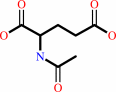 N-acetyl-L-glutamate
|
+
|
 ATP
ATP
|
=
|
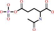 N-acetyl-L-glutamyl 5-phosphate
|
+
|
 ADP
ADP
|
|
|
|
|
|
|
|
|
|
|
|
|
Molecule diagrams generated from .mol files obtained from the
KEGG ftp site
|
|
|
|


 Links
Links


