 |
PDBsum entry 7e4x

|
|
|
|
|
|

|

|

|

|
|

|
|

|
|

|
|
|
 |

|

|
|
|
|
|
|
|
|
|
|
|
|
|
|
|
|
|
|
|
|
|
|
|
|
|
|
|
|
|
|
|
|
Metal binding protein
|
PDB id
|
|
|
|
7e4x
|
|
|
|
|
|
|
|
|
|
|
|
|
|
|

|

|

|
|
|
|
|
|
|
PDB id:
|
|
|
|
| Name: |
|
Metal binding protein
|
|
|
Title:
|
|
Structure of enolase from mycobacterium tuberculosis
|
|
Structure:
|
|
Enolase. Chain: a, b, c, d, e, f, g, h. Synonym: 2-phospho-d-glycerate hydro-lyase,2-phosphoglycerate dehydratase. Engineered: yes
|
|
Source:
|
|
Mycobacterium tuberculosis. Organism_taxid: 1773. Gene: eno, ayj03_005430, dsi38_18100, ers007663_02163, ers013471_01432, ers024276_01771, ers027659_01730, ers075361_03197, ers094182_03347, f6w99_03724, samea2683035_03102. Expressed in: mycolicibacterium smegmatis. Expression_system_taxid: 1772
|
|
Authors:
|
|
S.Bose,K.R.Vinothkumar
|
|
Key ref:
|
|
M.Ahmad
et al.
Structural snapshots of mycobacterium tuberculosis en reveal dual mode of 2pg binding and its implication i enzyme catalysis..
To be published,
.
PubMed id:
|
|
|
Date:
|
|
|
15-Feb-21
|
Release date:
|
16-Feb-22
|
|
|
|
|
|
|
PROCHECK
|

|
|
|
|
|
Headers
|
|
|
|
References
|
|
|
|
|
|

|
|
P9WNL1
(ENO_MYCTU) -
Enolase from Mycobacterium tuberculosis (strain ATCC 25618 / H37Rv)
|
|
|
|
Seq:
Struc:
|
|
|
|
429 a.a.
423 a.a.
|
|

|

|

|
|
|
|
|
|
|
|
|
|
|
Key: |
 |
PfamA domain |
|
|
 |
Secondary structure |
|
|
|
|
|
|
|
|
|
|
|
|
|
Enzyme class:
|
|
E.C.4.2.1.11
- phosphopyruvate hydratase.
|
|
|
|
|
|
|
Reaction:
|
|
(2R)-2-phosphoglycerate = phosphoenolpyruvate + H2O
|
|
|
|
|
|
(2R)-2-phosphoglycerate
|
=
|
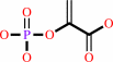 phosphoenolpyruvate
|
+
|
H2O
|
|
|
|
|
|
|
|
|
|
Cofactor:
|
|
Mg(2+)
|
|
|
|
|
|
|
|
|
Molecule diagrams generated from .mol files obtained from the
KEGG ftp site
|
|
|
|


 Links
Links


