 |
PDBsum entry 6dyt
 |
|
|
|
|
|
|
Enzyme class:
|
|
E.C.4.1.99.2
- tyrosine phenol-lyase.
|
|
|
|
|
|
|
Reaction:
|
|
L-tyrosine + H2O = phenol + pyruvate + NH4+
|
|
|
|
|
|
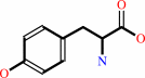 L-tyrosine
|
+
|
H2O
|
=
|
 phenol
|
+
|
 pyruvate
|
+
|
NH4(+)
|
|
|
|
|
|
|
|
|
|
Cofactor:
|
|
Pyridoxal 5'-phosphate
|
|
|
|
|
|
Pyridoxal 5'-phosphate
Bound ligand (Het Group name =
F0G)
matches with 68.18% similarity
|
|
|
|
|
|
|
Molecule diagrams generated from .mol files obtained from the
KEGG ftp site
|
|
|
|
|
|
|
|
|
|
|
|
|
|
 |
|
|
|
|
|
|
| |
|
|
| |
|
DOI no:
|
Biochemistry
57:6166-6179
(2018)
|
|
PubMed id:
|
|
|
|
|
|
| |
|
Crystal Structures of Wild-Type and F448A Mutant Citrobacter freundii Tyrosine Phenol-Lyase Complexed with a Substrate and Inhibitors: Implications for the Reaction Mechanism.
|
|
R.S.Phillips,
S.Craig.
|
|
|
|
|
| |
ABSTRACT
|
|
|
|
| |
|
|
Tyrosine phenol-lyase (TPL; EC 4.1.99.2) is a pyridoxal 5'-phosphate-dependent
enzyme that catalyzes the reversible hydrolytic cleavage of l-tyrosine to phenol
and ammonium pyruvate. We have shown previously that F448A TPL has
kcat and kcat/ Km values for l-tyrosine reduced
by ∼104-fold [Phillips, R. S., Vita, A., Spivey, J. B., Rudloff, A.
P., Driscoll, M. D., and Hay, S. (2016) ACS Catal. 6, 6770-6779]. We have now
obtained crystal structures of F448A TPL and complexes with l-alanine,
l-methionine, l-phenylalanine, and 3-F-l-tyrosine at 2.05-2.27 Å and the
complex of wild-type TPL with l-phenylalanine at 1.8 Å. The small domain of
F448A TPL, where Phe-448 is located, is more disordered in chain A than in
wild-type TPL. The complexes of F448A TPL with l-alanine and l-phenylalanine are
in an open conformation in both chains, while the complex with l-methionine is a
52:48 open:closed equilibrium mixture in chain A. Wild-type TPL with l-alanine
is closed in chain A and open in chain B, and the complex with l-phenylalanine
is a 56:44 open:closed mixture in chain A. Thus, the Phe-448 to alanine mutation
affects the conformational equilibrium of open and closed active sites. The
structure of the 3-F-l-tyrosine quinonoid complex of F448A TPL is unstrained and
in an open conformation, with a hydrogen bond from the phenolic OH to Thr-124.
These results support our previous conclusion that ground-state strain plays a
critical role in the mechanism of TPL.
|
|
|
|
|
|
|
|
|
|
|
|
|
|
|
|
|
|
 |
 |
 |
|


 Links
Links


