 |
PDBsum entry 4jm7

|
|
|
|
|
 |
Contents |
 |
|
|
|
|
|
|
|
|
|
|
|
 122 a.a.
|
|
|
|
|
|
|
|
|
|
|
 116 a.a.
|
|
|
|
|
|
|
|
|
|
|
 122 a.a.
|
|
|
|
|
|
|
|
|
|
|
|
|
PDB id:
|
|
|
|
| Name: |
|
Transferase
|
|
|
Title:
|
|
1.82 angstrom resolution crystal structure of holo-(acyl-carrier- protein) synthase (acps) from staphylococcus aureus
|
|
Structure:
|
|
Holo-[acyl-carrier-protein] synthase. Chain: a, b, c. Fragment: acyl-carrier-protein synthase. Synonym: holo-acp synthase, 4'-phosphopantetheinyl transferase acps. Engineered: yes
|
|
Source:
|
|
Staphylococcus aureus. Organism_taxid: 93062. Strain: col. Gene: acps, sacol2061. Expressed in: escherichia coli. Expression_system_taxid: 469008.
|
|
Resolution:
|
|
|
1.82Å
|
R-factor:
|
0.186
|
R-free:
|
0.225
|
|
|
Authors:
|
|
A.S.Halavaty,G.Minasov,L.Shuvalova,I.Dubrovska,L.Papazisi, W.F.Anderson,Center For Structural Genomics Of Infectious Diseases (Csgid)
|
|
Key ref:
|
|
A.S.Halavaty
et al.
(2012).
Structural characterization and comparison of three acyl-carrier-protein synthases from pathogenic bacteria.
Acta Crystallogr D Biol Crystallogr,
68,
1359-1370.
PubMed id:
|
|
|
Date:
|
|
|
13-Mar-13
|
Release date:
|
27-Mar-13
|
|
|
Supersedes:
|
|
|
|
|
|
|
|
PROCHECK
|

|
|
|
|
|
Headers
|
|
|
|
References
|
|
|
|
|
|

|
|
Q5HED0
(ACPS_STAAC) -
Holo-[acyl-carrier-protein] synthase from Staphylococcus aureus (strain COL)
|
|
|
|
Seq:
Struc:
|
|
|
|
119 a.a.
122 a.a.
|
|

|

|
|
|
|
|
|
|
|
|
|
|
|
|
|
|
|
|
|
Enzyme class:
|
|
Chains A, B, C:
E.C.2.7.8.7
- holo-[acyl-carrier-protein] synthase.
|
|
|
|
|
|
|
Reaction:
|
|
apo-[ACP] + CoA = holo-[ACP] + adenosine 3',5'-bisphosphate + H+
|
|
|
|
|
|
apo-[ACP]
|
+
|
 CoA
CoA
|
=
|
holo-[ACP]
|
+
|
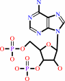 adenosine 3',5'-bisphosphate
|
+
|
H(+)
|
|
|
|
|
|
|
|
|
|
Cofactor:
|
|
Mg(2+)
|
|
|
|
|
|
|
|
|
Molecule diagrams generated from .mol files obtained from the
KEGG ftp site
|
|
|
|
|
|
|
|
|
|
|
|
|
|
 |
|
|
|
|
|
|
| |
|
|
| |
|
|
Acta Crystallogr D Biol Crystallogr
68:1359-1370
(2012)
|
|
PubMed id:
|
|
|
|
|
|
| |
|
Structural characterization and comparison of three acyl-carrier-protein synthases from pathogenic bacteria.
|
|
A.S.Halavaty,
Y.Kim,
G.Minasov,
L.Shuvalova,
I.Dubrovska,
J.Winsor,
M.Zhou,
O.Onopriyenko,
T.Skarina,
L.Papazisi,
K.Kwon,
S.N.Peterson,
A.Joachimiak,
A.Savchenko,
W.F.Anderson.
|
|
|
|
|
| |
ABSTRACT
|
|
|
|
| |
|
|
Some bacterial type II fatty-acid synthesis (FAS II) enzymes have been shown to
be important candidates for drug discovery. The scientific and medical quest for
new FAS II protein targets continues to stimulate research in this field. One of
the possible additional candidates is the acyl-carrier-protein synthase (AcpS)
enzyme. Its holo form post-translationally modifies the apo form of an acyl
carrier protein (ACP), which assures the constant delivery of thioester
intermediates to the discrete enzymes of FAS II. At the Center for Structural
Genomics of Infectious Diseases (CSGID), AcpSs from Staphylococcus aureus
(AcpS(SA)), Vibrio cholerae (AcpS(VC)) and Bacillus anthracis (AcpS(BA)) have
been structurally characterized in their apo, holo and product-bound forms,
respectively. The structure of AcpS(BA) is emphasized because of the two
3',5'-adenosine diphosphate (3',5'-ADP) product molecules that are found in each
of the three coenzyme A (CoA) binding sites of the trimeric protein. One
3',5'-ADP is bound as the 3',5'-ADP part of CoA in the known structures of the
CoA-AcpS and 3',5'-ADP-AcpS binary complexes. The position of the second
3',5'-ADP has never been described before. It is in close proximity to the first
3',5'-ADP and the ACP-binding site. The coordination of two ADPs in AcpS(BA) may
possibly be exploited for the design of AcpS inhibitors that can block binding
of both CoA and ACP.
|
|
|
|
|
|
|
|
|
|
|
|
|
|
|
|
|
|
 |
 |
 |
|
| |


 Links
Links


