 |
PDBsum entry 3loo

|
|
|
|
|
 |
Contents |
 |
|
|
|
|
|
|
|
|
|
|
|
|
|
|
* Residue conservation analysis
|
|
|
|
|
|
|
|
|
|
|
Enzyme class:
|
|
E.C.2.7.1.20
- adenosine kinase.
|
|
|
|
|
|
|
Reaction:
|
|
adenosine + ATP = AMP + ADP + H+
|
|
|
|
|
|
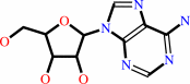 adenosine
|
+
|
ATP
Bound ligand (Het Group name = )
matches with 58.49% similarity
|
=
|
 AMP
AMP
|
+
|
 ADP
ADP
|
+
|
H(+)
|
|
|
|
|
|
|
|
|
|
|
|
|
Molecule diagrams generated from .mol files obtained from the
KEGG ftp site
|
|
|
|
|
|
|
|
|
|
|
|
|
|
 |
|
|
|
|
|
|
| |
|
|
| |
|
|
Biochemistry
50:1885-1893
(2011)
|
|
PubMed id:
|
|
|
|
|
|
| |
|
A high-affinity adenosine kinase from Anopheles gambiae.
|
|
M.B.Cassera,
M.C.Ho,
E.F.Merino,
E.S.Burgos,
A.Rinaldo-Matthis,
S.C.Almo,
V.L.Schramm.
|
|
|
|
|
| |
ABSTRACT
|
|
|
|
| |
|
|
Genome analysis revealed a mosquito orthologue of adenosine kinase in Anopheles
gambiae (AgAK; the most important vector for the transmission of Plasmodium
falciparum in Africa). P. falciparum are purine auxotrophs and do not express an
adenosine kinase but rely on their hosts for purines. AgAK was kinetically
characterized and found to have the highest affinity for adenosine (K(m) = 8.1
nM) of any known adenosine kinase. AgAK is specific for adenosine at the
nucleoside site, but several nucleotide triphosphate phosphoryl donors are
tolerated. The AgAK crystal structure with a bound bisubstrate analogue Ap(4)A
(2.0 Å resolution) reveals interactions for adenosine and ATP and the geometry
for phosphoryl transfer. The polyphosphate charge is partly neutralized by a
bound Mg(2+) ion and an ion pair to a catalytic site Arg. The AgAK structure
consists of a large catalytic core in a three-layer α/β/α sandwich, and a
small cap domain in contact with adenosine. The specificity and tight binding
for adenosine arise from hydrogen bond interactions of Asn14, Leu16, Leu40,
Leu133, Leu168, Phe168, and Thr171 and the backbone of Ile39 and Phe168 with the
adenine ring as well as through hydrogen bond interactions between Asp18, Gly64,
and Asn68 and the ribosyl 2'- and 3'-hydroxyl groups. The structure is more
similar to that of human adenosine kinase (48% identical) than to that of AK
from Toxoplasma gondii (31% identical). With this extraordinary affinity for
AgAK, adenosine is efficiently captured and converted to AMP at near the
diffusion limit, suggesting an important role for this enzyme in the maintenance
of the adenine nucleotide pool. mRNA analysis verifies that AgAK transcripts are
produced in the adult insects.
|
|
|
|
|
|
|
|
|
|
|
|
|
|
|
|
|
|
 |
 |
 |
|


 Links
Links


