 |
PDBsum entry 3c3i

|
|
|
|
|
 |
Contents |
 |
|
|
|
|
|
|
|
|
|
|
|
|
|
|
* Residue conservation analysis
|
|
|
|
|
|
|
|
|
|
Enzyme class:
|
|
Chains A, B, C, D:
E.C.3.6.1.23
- dUTP diphosphatase.
|
|
|
|
|
|
|
Reaction:
|
|
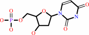
dUTP + H2O = dUMP + diphosphate + H+
|
|
|
|
|
|
dUTP
Bound ligand (Het Group name = )
matches with 85.71% similarity
|
+
|
H2O
|
=
|
 dUMP
dUMP
|
+
|
 diphosphate
|
+
|
H(+)
|
|
|
|
|
|
|
|
|
|
|
|
|
Molecule diagrams generated from .mol files obtained from the
KEGG ftp site
|
|
|
|
|
|
|
|
|
|
|
|
|
|
 |
|
|
|
|
|
|
| |
|
|
| |
|
|
Acta Crystallogr Sect F Struct Biol Cryst Commun
65:1030-1034
(2009)
|
|
PubMed id:
|
|
|
|
|
|
| |
|
Crystallization and crystal-packing studies of Chlorella virus deoxyuridine triphosphatase.
|
|
K.Homma,
H.Moriyama.
|
|
|
|
|
| |
ABSTRACT
|
|
|
|
| |
|
|
The 141-amino-acid deoxyuridine triphosphatase (dUTPase) from the algal
Chlorella virus IL-3A and its Glu81Ser/Thr84Arg-mutant derivative Mu-22 were
crystallized using the hanging-drop vapor-diffusion method at 298 K with
polyethylene glycol as the precipitant. An apo IL-3A dUTPase with an
amino-terminal T7 epitope tag and a carboxy-terminal histidine tag yielded cubic
P2(1)3 crystals with unit-cell parameter a = 106.65 A. In the presence of dUDP,
the enzyme produced thin stacked orthorhombic P222 crystals with unit-cell
parameters a = 81.0, b = 96.2, c = 132.8 A. T7-histidine-tagged Mu-22 dUTPase
formed thin stacked rectangular crystals. Amino-terminal histidine-tagged
dUTPases did not crystallize but formed aggregates. Glycyl-seryl-tagged dUTPases
yielded cubic P2(1)3 IL-3A crystals with unit-cell parameter a = 105.68 A and
hexagonal P6(3) Mu-22 crystals with unit-cell parameters a = 132.07, c = 53.45
A, gamma = 120 degrees . Owing to the Thr84Arg mutation, Mu-22 dUTPase had
different monomer-to-monomer interactions to those of IL-3A dUTPase.
|
|
|
|
|
|
|
|
|
|
|
|
|
|
|
|
|
|
 |
 |
 |
|
|


 Links
Links


