 |
PDBsum entry 2d69
|
|

|
|
PDB id:
|
|
|
|
| Name: |
|
Lyase
|
|
|
Title:
|
|
Crystal structure of the complex of sulfate ion and octameric ribulose-1,5-bisphosphate carboxylase/oxygenase (rubisco) from pyrococcus horikoshii ot3 (form-2 crystal)
|
|
Structure:
|
|
Ribulose bisphosphate carboxylase. Chain: a, b, d, e. Synonym: rubisco, ribulose-1,5-bisphosphate carboxylase/oxygenase. Engineered: yes
|
|
Source:
|
|
Pyrococcus horikoshii. Organism_taxid: 70601. Strain: ot3. Expressed in: escherichia coli bl21(de3). Expression_system_taxid: 469008.
|
|
Biol. unit:
|
|
 Dimer (from PDB file)
|
|
Resolution:
|
|
|
1.90Å
|
R-factor:
|
0.181
|
R-free:
|
0.209
|
|
|
Authors:
|
|
E.Mizohata,C.Mishima,R.Akasaka,H.Uda,T.Terada,M.Shirouzu,S.Yokoyama, Riken Structural Genomics/proteomics Initiative (Rsgi)
|
|
Key ref:
|
|
E.Mizohata
et al.
Crystal structure of the complex of sulfate ion and octameric ribulose-1,5-Bisphosphate carboxylase/oxygenase (rubisco) from pyrococcus horikoshii ot3 (form-2 crystal).
To be published,
.
|
|
|
Date:
|
|
|
10-Nov-05
|
Release date:
|
10-May-06
|
|
|
|
|
|
|
PROCHECK
|

|
|
|
|
|
Headers
|
|
|
|
References
|
|
|
|
|
|

|
|
O58677
(RBL_PYRHO) -
Ribulose bisphosphate carboxylase from Pyrococcus horikoshii (strain ATCC 700860 / DSM 12428 / JCM 9974 / NBRC 100139 / OT-3)
|
|
|
|
Seq:
Struc:
|
|
|
|
430 a.a.
424 a.a.
|
|

|

|

|
|
|
|
|
|
|
|
|
|
|
Key: |
 |
PfamA domain |
|
|
 |
Secondary structure |
|
 |
CATH domain |
|
|
|
|
|
|
|
|
|
|
|
|
|
Enzyme class:
|
|
E.C.4.1.1.39
- ribulose-bisphosphate carboxylase.
|
|
|
|
|
|
|
Reaction:
|
|
2 (2R)-3-phosphoglycerate + 2 H+ = D-ribulose 1,5-bisphosphate + CO2 + H2O
|
|
|
|
|
|
2
×
(2R)-3-phosphoglycerate
|
+
|
2
×
H(+)
|
=
|
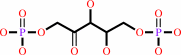 D-ribulose 1,5-bisphosphate
|
+
|
 CO2
CO2
|
+
|
H2O
|
|
|
|
|
|
|
|
|
|
|
|
|
Molecule diagrams generated from .mol files obtained from the
KEGG ftp site
|
|
|
|


 Links
Links


