 |
PDBsum entry 2b7n

|
|
|
|
|
 |
Contents |
 |
|
|
|
|
|
|
|
|
|
|
|
|
* Residue conservation analysis
|
|
|
|
|
|
|
|
|
|
|
Enzyme class:
|
|
E.C.2.4.2.19
- nicotinate-nucleotide diphosphorylase (carboxylating).
|
|
|
|
|
|
|
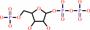
Reaction:
|
|
nicotinate beta-D-ribonucleotide + CO2 + diphosphate = quinolinate + 5-phospho-alpha-D-ribose 1-diphosphate + 2 H+
|
|
|
|
|
|
nicotinate beta-D-ribonucleotide
|
+
|
 CO2
CO2
|
+
|
 diphosphate
|
=
|
 quinolinate
|
+
|
 5-phospho-alpha-D-ribose 1-diphosphate
|
+
|
2
×
H(+)
Bound ligand (Het Group name = )
corresponds exactly
|
|
|
|
|
|
|
|
|
|
|
|
|
Molecule diagrams generated from .mol files obtained from the
KEGG ftp site
|
|
|
|
|
|
|
|
|
|
|
|
|
|
 |
|
|
|
|
|
|
| |
|
|
| |
|
DOI no:
|
Proteins
63:252-255
(2006)
|
|
PubMed id:
|
|
|
|
|
|
| |
|
Crystal structure of quinolinic acid phosphoribosyltransferase from Helicobacter pylori.
|
|
M.K.Kim,
Y.J.Im,
J.H.Lee,
S.H.Eom.
|
|
|
|
|
| |
ABSTRACT
|
|
|
|
| |
|
|
|
|
| |
Selected figure(s)
|
|
|
|
| |
|
|

|
|
Figure 1.
Figure 1. Crystal structure of Hp-QAPRTase. (A) Ribbon diagram
of the Hp-QAPRTase monomer. The N-terminal domain (residues
1-116, 258-273) is shown in yellow, and the C-terminal domain
(residue 117-257) in orange. QA is shown as a space filling
model. (B) Structure of the Hp-QAPRTase dimer. (C) QA binding
site. The side chains at the active site are shown as a
ball-and-stick model. (D) NAMN-binding site. The 2Fo-Fc electron
density map contoured at 1s. (E) Structure of the Hp-QAPRTase
hexamer. (F). Surface representation of the Hp-QAPRTase hexamer.
The side chains of Phe181 at the interface of the subunits are
shown as a space filling model. All figures were prepared using
the program PyMOL (www.pymol.org).
|
|
|
|
|
|
| |
The above figure is
reprinted
by permission from John Wiley & Sons, Inc.:
Proteins
(2006,
63,
252-255)
copyright 2006.
|
|
|
|
|
|
|
|
|
|
|
|
|
|
 |
 |
 |
|
Literature references that cite this PDB file's key reference
|
|
|
| |
PubMed id
|
|
Reference
|
|
|
|
|
|
Z.Bello,
and
C.Grubmeyer
(2010).
Roles for cationic residues at the quinolinic acid binding site of quinolinate phosphoribosyltransferase.
|
| |
Biochemistry,
49,
1388-1395.
|
|
|
|
|
|
|
M.K.Kim,
G.B.Kang,
W.K.Song,
and
S.H.Eom
(2007).
The role of Phe181 in the hexamerization of Helicobacter pylori quinolinate phosphoribosyltransferase.
|
| |
Protein J,
26,
517-521.
|
|
|
|
|
|
The most recent references are shown first.
Citation data come partly from CiteXplore and partly
from an automated harvesting procedure. Note that this is likely to be
only a partial list as not all journals are covered by
either method. However, we are continually building up the citation data
so more and more references will be included with time.
|
|


 Links
Links


